Instituto de Tecnologia Química e Biológica António Xavier, Universidade Nova de Lisboa, Oeiras, Portugal.
Instituto de Biologia Experimental e Tecnológica, Oeiras, Portugal.
Sci Rep. 2019 Sep 20;9(1):13615. doi: 10.1038/s41598-019-49944-x.
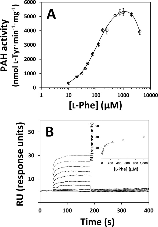
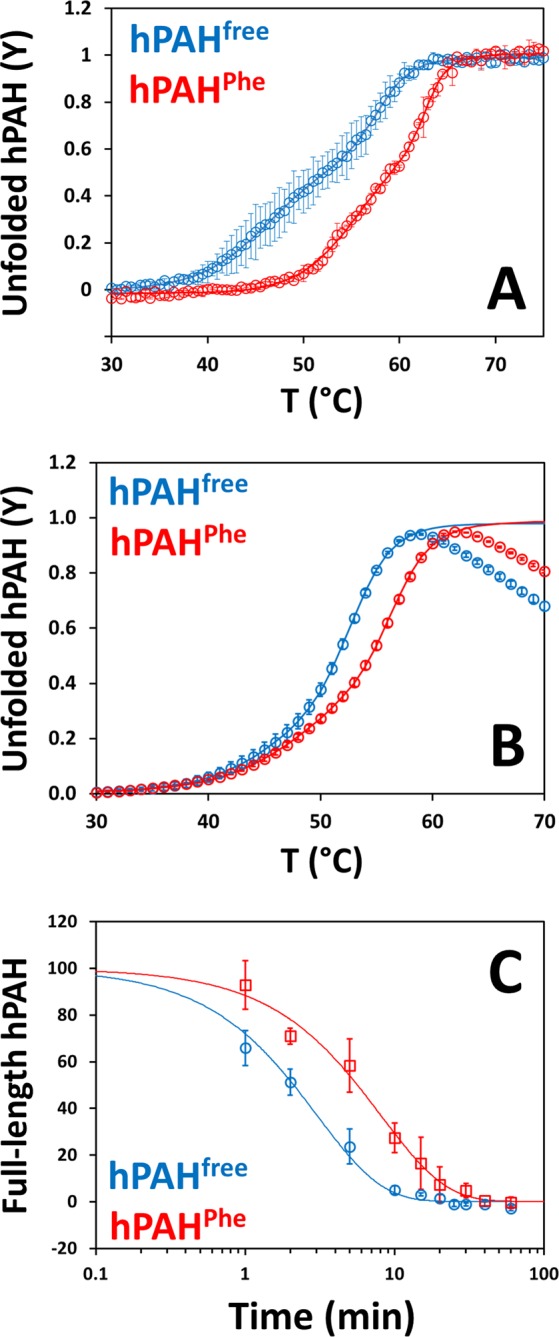
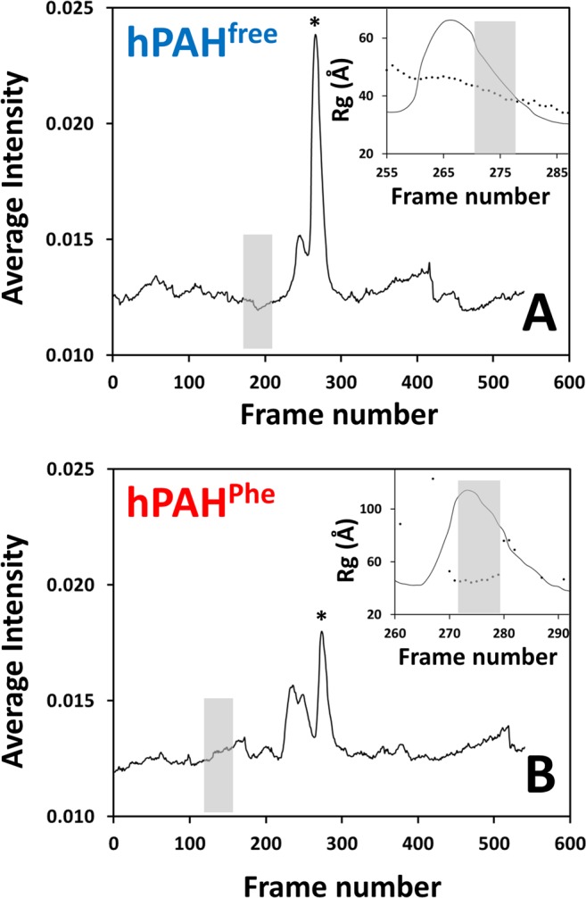
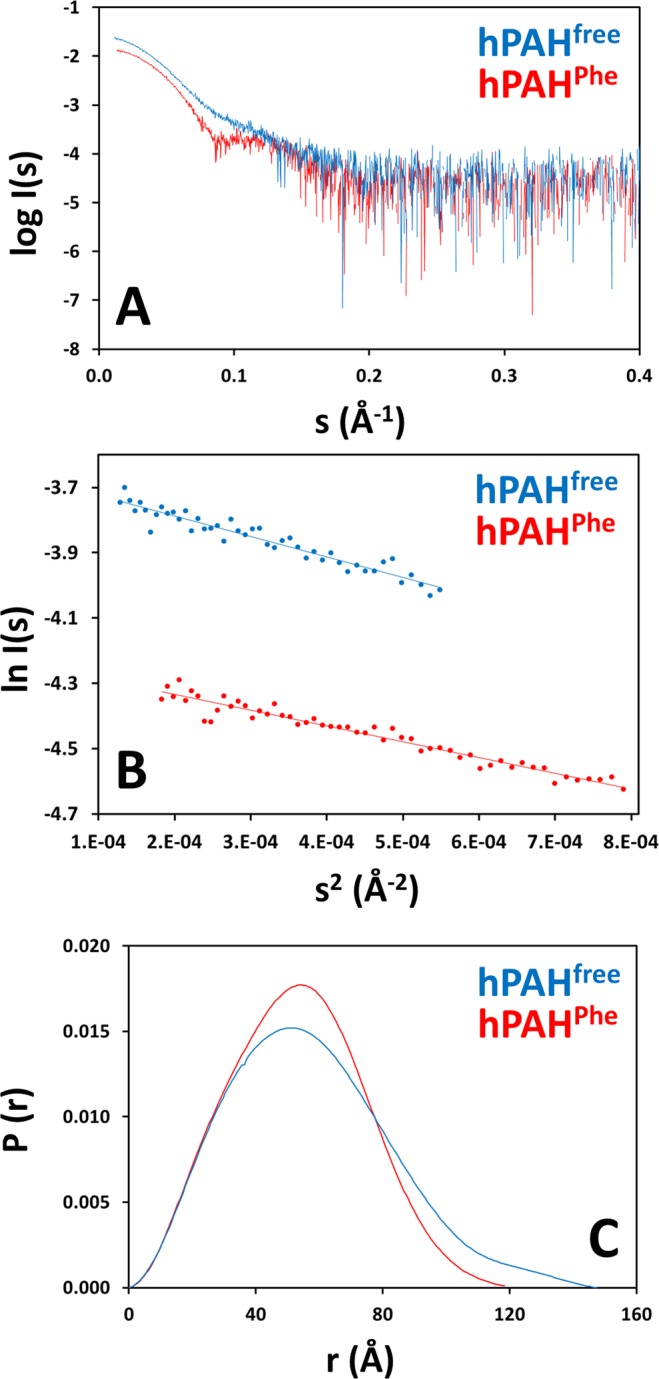
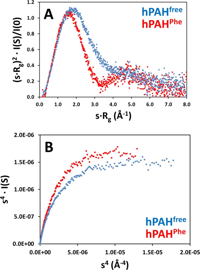
Human phenylalanine hydroxylase (hPAH) hydroxylates L-phenylalanine (L-Phe) to L-tyrosine, a precursor for neurotransmitter biosynthesis. Phenylketonuria (PKU), caused by mutations in PAH that impair PAH function, leads to neurological impairment when untreated. Understanding the hPAH structural and regulatory properties is essential to outline PKU pathophysiological mechanisms. Each hPAH monomer comprises an N-terminal regulatory, a central catalytic and a C-terminal oligomerisation domain. To maintain physiological L-Phe levels, hPAH employs complex regulatory mechanisms. Resting PAH adopts an auto-inhibited conformation where regulatory domains block access to the active site. L-Phe-mediated allosteric activation induces a repositioning of the regulatory domains. Since a structure of activated wild-type hPAH is lacking, we addressed hPAH L-Phe-mediated conformational changes and report the first solution structure of the allosterically activated state. Our solution structures obtained by small-angle X-ray scattering support a tetramer with distorted P222 symmetry, where catalytic and oligomerisation domains form a core from which regulatory domains protrude, positioning themselves close to the active site entrance in the absence of L-Phe. Binding of L-Phe induces a large movement and dimerisation of regulatory domains, exposing the active site. Activated hPAH is more resistant to proteolytic cleavage and thermal denaturation, suggesting that the association of regulatory domains stabilises hPAH.
人苯丙氨酸羟化酶(hPAH)将 L-苯丙氨酸(L-Phe)羟化为 L-酪氨酸,这是神经递质生物合成的前体。苯丙酮尿症(PKU)是由 PAH 基因突变导致 PAH 功能受损引起的,如果不治疗,会导致神经损伤。了解 hPAH 的结构和调节特性对于概述 PKU 的病理生理机制至关重要。每个 hPAH 单体由 N 端调节域、中央催化域和 C 端寡聚化域组成。为了维持生理 L-Phe 水平,hPAH 采用复杂的调节机制。静息状态下的 PAH 采用自动抑制构象,其中调节域阻止进入活性部位。L-Phe 介导的别构激活诱导调节域的重新定位。由于缺乏激活的野生型 hPAH 结构,我们研究了 hPAH 与 L-Phe 介导的构象变化,并报告了第一个别构激活状态的溶液结构。我们通过小角 X 射线散射获得的溶液结构支持四聚体具有扭曲的 P222 对称性,其中催化和寡聚化域形成一个核心,调节域从该核心伸出,在没有 L-Phe 的情况下,它们靠近活性部位入口定位。L-Phe 的结合诱导调节域的大运动和二聚化,暴露出活性部位。激活的 hPAH 对蛋白水解切割和热变性更具抗性,这表明调节域的缔合稳定了 hPAH。