Department of Acupuncture‑Moxibustion and Tuina, The Third Affiliated Hospital of Chengdu University of Traditional Chinese Medicine, Chengdu, Sichuan 611137, P.R. China.
Yueyang Hospital of Integrated Traditional Chinese and Western Medicine, Shanghai University of Traditional Chinese Medicine, Shanghai 200437, P.R. China.
Mol Med Rep. 2019 Nov;20(5):4101-4110. doi: 10.3892/mmr.2019.10674. Epub 2019 Sep 11.
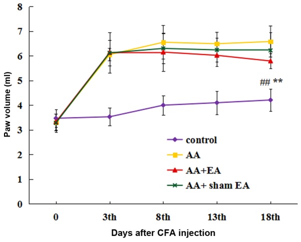
p53 and mouse double minute 2 homolog (MDM2) serve key regulatory roles in the apoptosis of synovial cells. The present study aimed to investigate the effects of electroacupuncture (EA) at the 'Zusanli' (ST36) and 'Xuanzhong' (GB39) acupoints on apoptosis in an adjuvant arthritis (AA) rat model. A total of 40 male Sprague‑Dawley rats were randomly divided into Control, AA, AA + EA and AA + sham EA groups (n=10 rats in each group). Rats in all the groups, with the exception of the control group, were injected with Complete™ Freund's adjuvant into the bilateral hindlimb footpad to establish the AA model. Rats in the AA + EA group were treated with EA at the ST36 and GB39 acupoints. Rats in the AA + sham EA group were treated with percutaneous electrical stimulation at a position of 5 mm away from the ST36 and GB39 acupoints. The arthritis index scores and hindlimb paw volumes of the rats in each group were recorded. Subsequently, pathological changes in the synovial tissue were evaluated by hematoxylin and eosin (H&E) staining, and the apoptotic rate of the synovial cells was detected by TUNEL staining. In addition, the expression levels of the apoptosis‑associated proteins, Bax, phorbol‑12‑myristate‑13‑acetate‑induced protein 1 (Noxa) and p53 upregulated modulator of apoptosis (PUMA), were determined by western blot analysis. The expression of both the gene and protein of p53 and MDM2 in synovial tissue was detected by reverse transcription‑quantitative polymerase chain reaction (RT‑qPCR) and western blot analysis, respectively. The results indicated that the arthritis index scores and hindlimb paw volumes upon EA stimulation were significantly decreased compared with those of the AA group (P<0.05). H&E staining revealed that the synovial inflammation of EA stimulation was significantly decreased compared with the AA group (P<0.05). The TUNEL assay results indicated that the apoptotic rate of synovial cells in the AA + EA group was significantly increased compared with that in the AA group (P<0.05). Furthermore, an increased expression of proapoptotic proteins was confirmed by the increased expression levels of Bax, Noxa and PUMA in the AA + EA group. The results of RT‑qPCR and western blot analysis demonstrated that, compared with the AA group, EA stimulation led to a marked increase in p53 (P<0.05) and a significant decrease in MDM2 (P<0.05) gene and protein expression. Taken together, these results demonstrated that EA performed on the ST36 and GB39 acupoints led to a significant amelioration in AA injury of model rats, by regulating the p53 signaling pathway and inducing apoptosis.
p53 和鼠双微体 2 同源物(MDM2)在滑膜细胞凋亡中起关键调节作用。本研究旨在探讨电针对佐剂性关节炎(AA)大鼠模型滑膜细胞凋亡的影响。
将 40 只雄性 Sprague-Dawley 大鼠随机分为对照组、AA 组、AA+EA 组和 AA+假电针(sham EA)组(每组 10 只大鼠)。除对照组外,所有组大鼠均在双侧后肢足底注射完全弗氏佐剂以建立 AA 模型。AA+EA 组大鼠接受 ST36 和 GB39 穴位电针治疗。AA+sham EA 组大鼠在 ST36 和 GB39 穴位 5mm 处接受经皮电刺激治疗。记录各组大鼠关节炎指数评分和后肢爪体积。随后,通过苏木精和伊红(H&E)染色评估滑膜组织的病理变化,通过 TUNEL 染色检测滑膜细胞的凋亡率。此外,通过 Western blot 分析测定凋亡相关蛋白 Bax、佛波醇 12-肉豆蔻酸 13-乙酸酯诱导蛋白 1(Noxa)和 p53 上调凋亡调节剂(PUMA)的表达水平。通过逆转录-定量聚合酶链反应(RT-qPCR)和 Western blot 分析分别检测滑膜组织中 p53 和 MDM2 的基因和蛋白表达。
结果表明,与 AA 组相比,电针刺激后的关节炎指数评分和后肢爪体积显著降低(P<0.05)。H&E 染色显示,电针刺激后的滑膜炎症明显低于 AA 组(P<0.05)。TUNEL 检测结果表明,AA+EA 组滑膜细胞的凋亡率明显高于 AA 组(P<0.05)。此外,通过 Bax、Noxa 和 PUMA 表达水平的增加,证实了促凋亡蛋白的表达增加。RT-qPCR 和 Western blot 分析结果表明,与 AA 组相比,电针刺激导致 p53 显著增加(P<0.05),MDM2 基因和蛋白表达显著降低(P<0.05)。
综上所述,电针 ST36 和 GB39 穴位可显著改善 AA 损伤模型大鼠的 AA 损伤,其机制可能与调节 p53 信号通路和诱导细胞凋亡有关。