Zhu Jun, Su Chengguo, Chen Yuzhou, Hao Xinyu, Jiang Jianzhen
School of Acupuncture-Moxibustion and Tuina, The Third Affiliated Hospital, Chengdu University of Traditional Chinese Medicine, Chengdu 610075, China.
Evid Based Complement Alternat Med. 2019 Jun 16;2019:5741931. doi: 10.1155/2019/5741931. eCollection 2019.
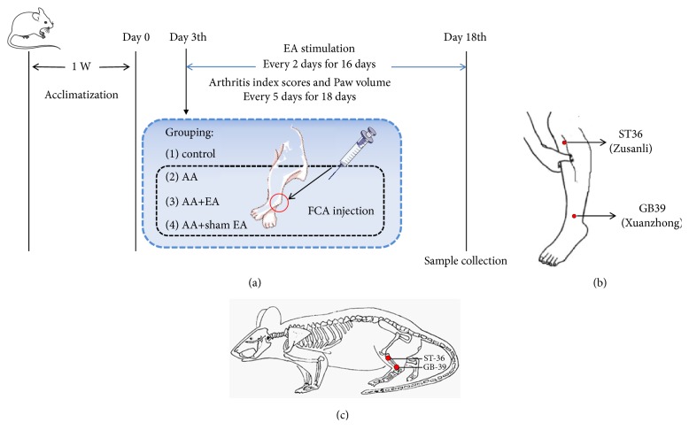
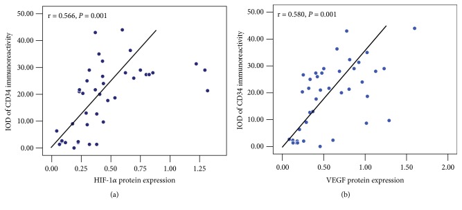
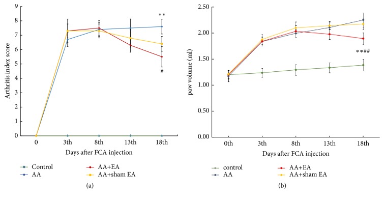
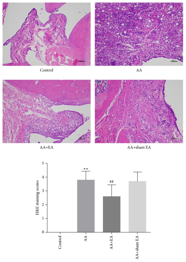
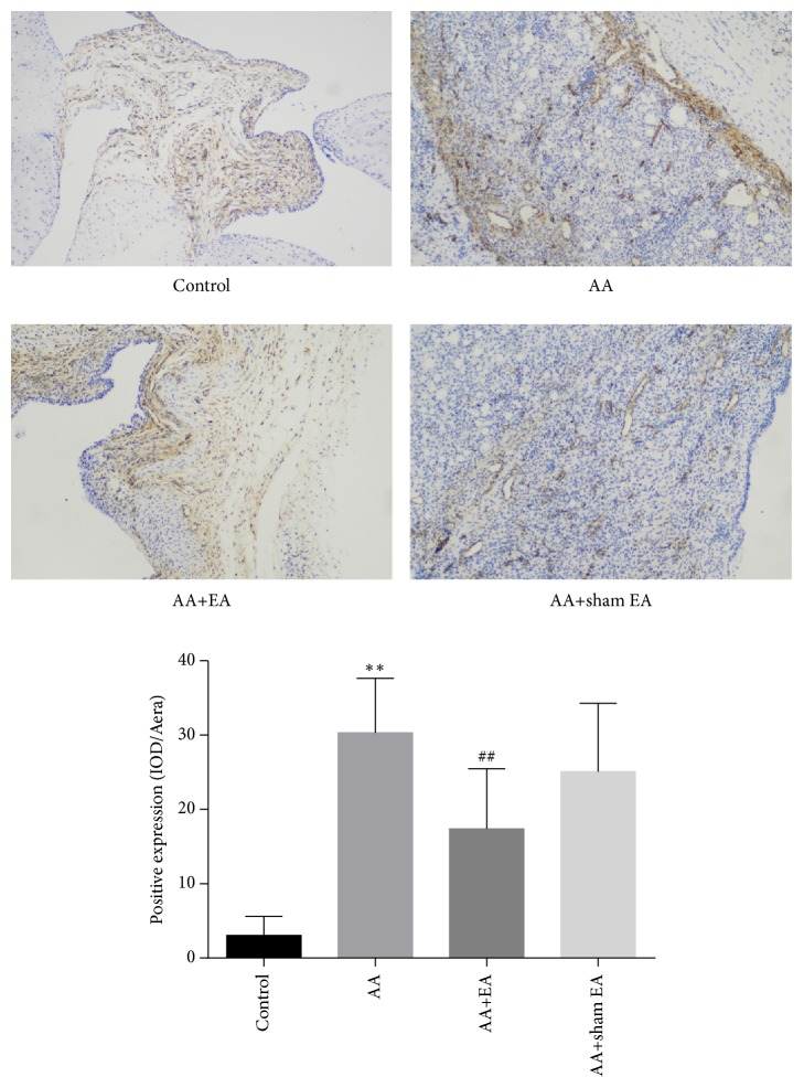
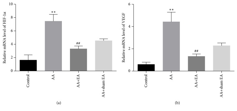
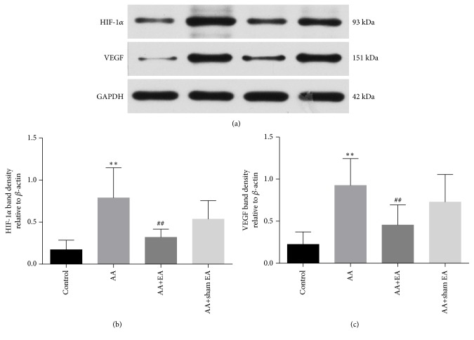
The hypoxia inducible factor-1 (HIF-1) and vascular endothelial growth factor (VEGF) play a key role in synovial angiogenesis in rheumatoid arthritis (RA). Therefore, this study aimed to test the hypothesis that electroacupuncture (EA) may inhibit RA synovial angiogenesis via HIF-1/VEGF expression. Sprague-Dawley rats were randomly distributed to 4 groups: control, adjuvant arthritis (AA), AA+electroacupuncture (AA+EA), and AA+sham EA groups. AA model was induced by injection of Freund's complete adjuvant in bilateral hind footpad. 3 days after injection, EA was delivered to the acupoints Zusanli (ST 36) and Xuanzhong (GB 39) once every two days for a total of 8 times in the AA+EA group, while sham EA treatment was applied in the AA+sham EA group. The arthritis score, paw volume, and H&E staining for each animal were measured. CD34 expression in synovial tissue of ankle joint was observed by immunohistochemistry. HIF-1 and VEGF mRNA and protein levels in synovial tissue were determined by real-time quantitative PCR and Western blot, respectively. Compared with rats in AA group, EA stimulation significantly decreased arthritis scores, paw volume, and pathological damage of synovial tissues. Moreover, EA markedly suppressed the synovial angiogenesis of AA rats, as evidenced by reduced CD34 positive expression. Furthermore, EA significantly reduced HIF-1 and VEGF mRNA and protein levels in synovial of AA rats. Finally, the CD34 expression in synovial tissue was positively correlated with HIF-1 and VEGF protein levels. . EA on ST36 and GB39 acupoints can effectively inhibit synovial angiogenesis in the AA rat model via downregulating HIF-1/VEGF expression.
缺氧诱导因子-1(HIF-1)和血管内皮生长因子(VEGF)在类风湿关节炎(RA)的滑膜血管生成中起关键作用。因此,本研究旨在验证电针(EA)可能通过HIF-1/VEGF表达抑制RA滑膜血管生成这一假说。将Sprague-Dawley大鼠随机分为4组:对照组、佐剂性关节炎(AA)组、AA+电针(AA+EA)组和AA+假电针组。通过在双侧后足垫注射弗氏完全佐剂诱导AA模型。注射后3天,AA+EA组每两天对足三里(ST 36)和悬钟(GB 39)穴位进行一次电针治疗,共8次,而AA+假电针组进行假电针治疗。测量每只动物的关节炎评分、爪体积,并进行苏木精-伊红(H&E)染色。通过免疫组织化学观察踝关节滑膜组织中CD34的表达。分别通过实时定量PCR和蛋白质印迹法测定滑膜组织中HIF-1和VEGF的mRNA和蛋白质水平。与AA组大鼠相比,电针刺激显著降低了关节炎评分、爪体积和滑膜组织的病理损伤。此外,电针明显抑制了AA大鼠的滑膜血管生成,CD34阳性表达减少证明了这一点。此外,电针显著降低了AA大鼠滑膜中HIF-1和VEGF的mRNA和蛋白质水平。最后,滑膜组织中CD34的表达与HIF-1和VEGF蛋白水平呈正相关。针刺ST36和GB39穴位的电针可通过下调HIF-1/VEGF表达有效抑制AA大鼠模型中的滑膜血管生成。