Department of Anatomy, College of Korean Medicine, Dongguk University, Gyeongju, Gyeongbuk 38066, Korea.
Department of Biomedical Science and Research Institute for Bioscience and Biotechnology, Hallym University, Chuncheon, Gangwon 24252, Korea.
Cells. 2019 Sep 22;8(10):1126. doi: 10.3390/cells8101126.
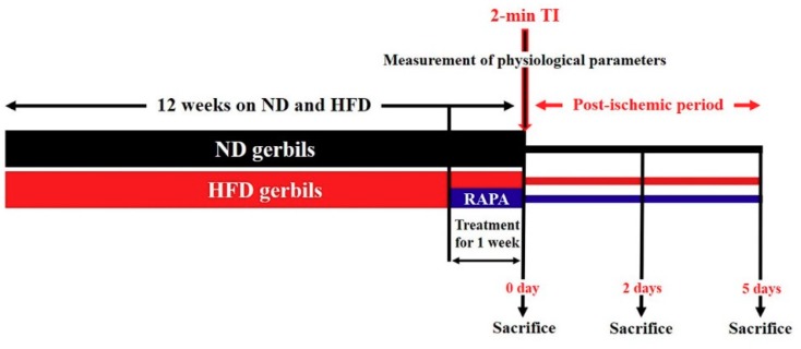
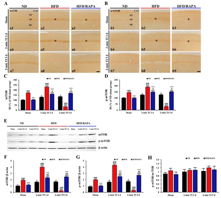
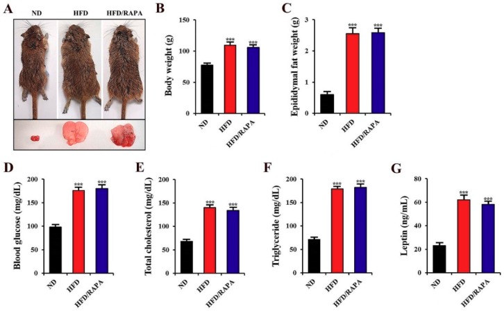
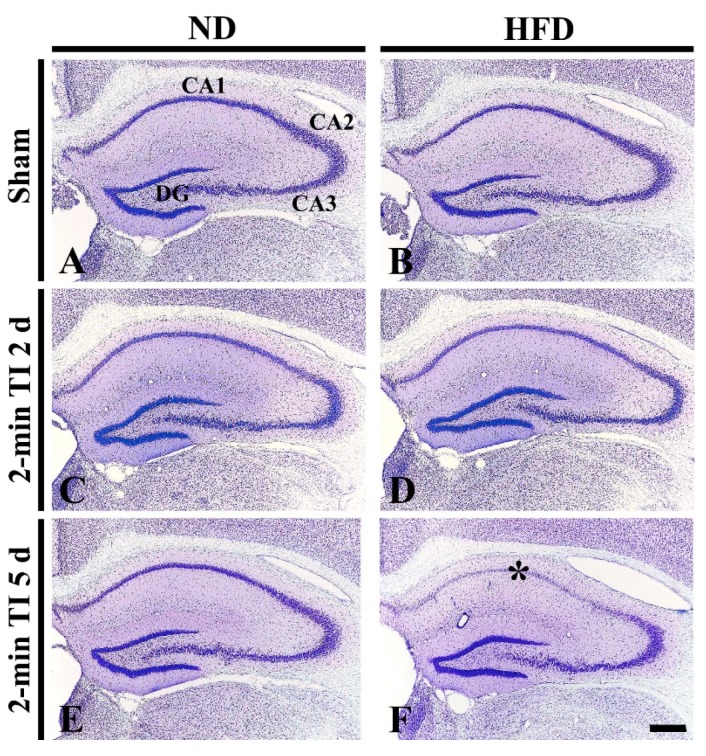
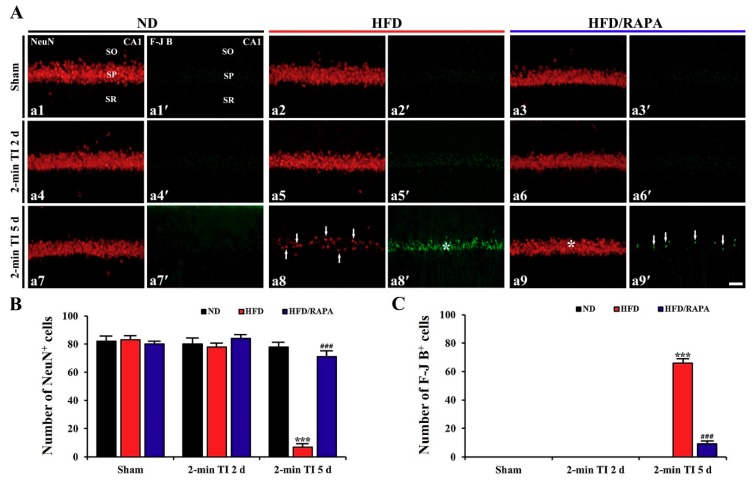
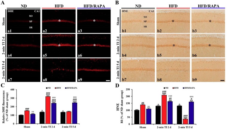
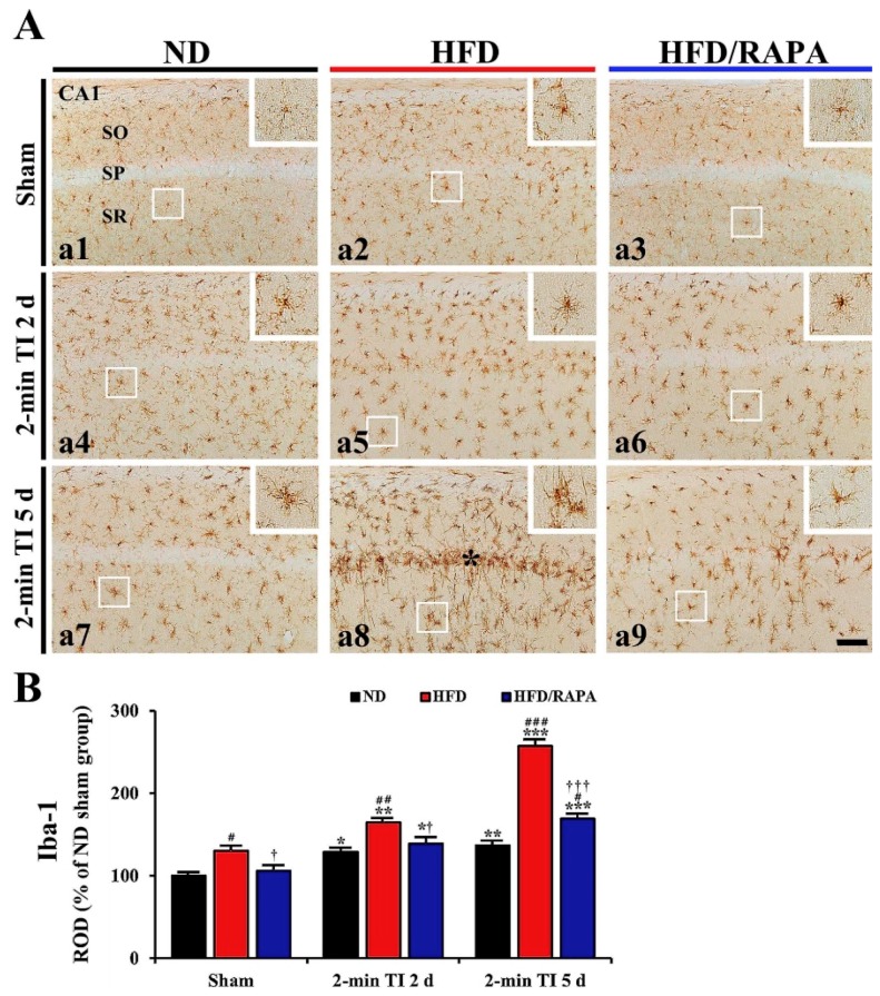
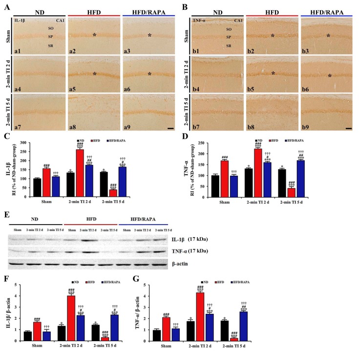
A brief episode of transient ischemia (TI) can confer cerebral ischemic tolerance against a subsequent severer TI under standard condition. The brain under obesity's conditions is more sensitive to ischemic injury. However, the impact of a brief episode of TI under obesity's conditions has not been fully addressed yet. Thus, the objective of this study was to determine the effect of a brief TI in the hippocampus of high-fat diet (HFD)-induced obese gerbils and related mechanisms. Gerbils were maintained on HFD or normal diet (ND) for 12 weeks and subjected to 2 min TI. HFD gerbils were heavier, with higher blood glucose, serum total cholesterol, triglycerides, and leptin levels. Massive loss of pyramidal neurons occurred in the hippocampal cornu ammonis 1 (CA1) field of HFD animals at 5 days after 2 min of TI, but 2 min of TI did not elicit death of pyramidal neurons in ND gerbils. The HFD group showed significantly increased levels of oxidative stress indicators (dihydroethidium and 4-hydroxynonenal) and proinflammatory cytokines (tumor necrosis factor-α and interleukin-1β) and microglial activation in pre- and/or post-ischemic phases compared to the ND group. Levels of mammalian target of rapamycin (mTOR) and phosphorylated-mTOR in the CA1 field of the HFD group were also significantly higher than the ND group. On the other hand, inhibition of mTOR activation by rapamycin (an allosteric mTOR inhibitor) significantly attenuated neuronal death induced by HFD, showing reduction of HFD-induced increases of oxidative stress indicators and proinflammatory cytokines, and microglia activation. Taken together, a brief episode of TI can evoke neuronal death under obesity's conditions. It might be closely associated with an abnormal increase of mTOR activation-mediated, severe oxidative stress and neuroinflammation in pre- and/or post-ischemic phases.
短暂性脑缺血发作(TI)可在标准条件下引起随后更严重的 TI 产生脑缺血耐受。肥胖状态下的大脑对缺血性损伤更为敏感。然而,肥胖状态下短暂性脑缺血发作的影响尚未得到充分研究。因此,本研究旨在确定短暂性脑缺血发作对高脂肪饮食(HFD)诱导肥胖沙鼠海马的影响及其相关机制。沙鼠维持在 HFD 或正常饮食(ND) 12 周后进行 2 分钟 TI。HFD 沙鼠体重增加,血糖、血清总胆固醇、甘油三酯和瘦素水平升高。2 分钟 TI 后 5 天,HFD 动物海马角 1(CA1)区大量锥体神经元丢失,但 ND 沙鼠 2 分钟 TI 不会引起锥体神经元死亡。与 ND 组相比,HFD 组在缺血前和/或缺血后阶段显示出明显增加的氧化应激标志物(二氢乙啶和 4-羟壬烯醛)和促炎细胞因子(肿瘤坏死因子-α和白细胞介素-1β)以及小胶质细胞激活水平。HFD 组 CA1 区哺乳动物雷帕霉素靶蛋白(mTOR)和磷酸化-mTOR 的水平也明显高于 ND 组。另一方面,雷帕霉素(一种变构 mTOR 抑制剂)抑制 mTOR 激活可显著减轻 HFD 诱导的神经元死亡,显示出降低 HFD 诱导的氧化应激标志物和促炎细胞因子以及小胶质细胞激活的增加。总之,短暂性脑缺血发作可在肥胖状态下引起神经元死亡。这可能与缺血前和/或缺血后阶段 mTOR 激活介导的异常增加、严重氧化应激和神经炎症密切相关。