Lin Xin, Li Gen, Zhang Yu, Zhao Jingjing, Lu Jiawen, Gao Yunge, Liu Huihui, Li Geng-Lin, Yang Tao, Song Lei, Wu Hao
Department of Otolaryngology-Head and Neck Surgery, Shanghai Ninth People's Hospital, Shanghai Jiao Tong University School of Medicine, Shanghai 200011, China.
Ear Institute, Shanghai Jiao Tong University School of Medicine, Shanghai 200125, China.
Aging (Albany NY). 2019 Sep 27;11(18):7416-7441. doi: 10.18632/aging.102246.
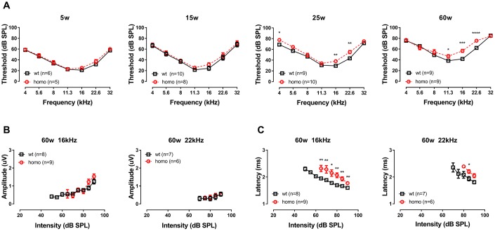
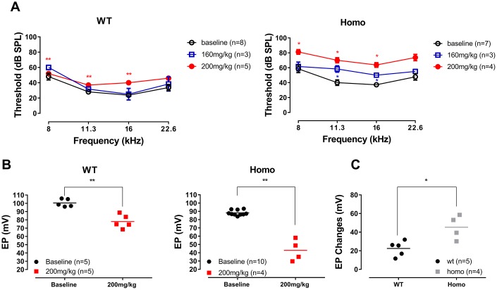
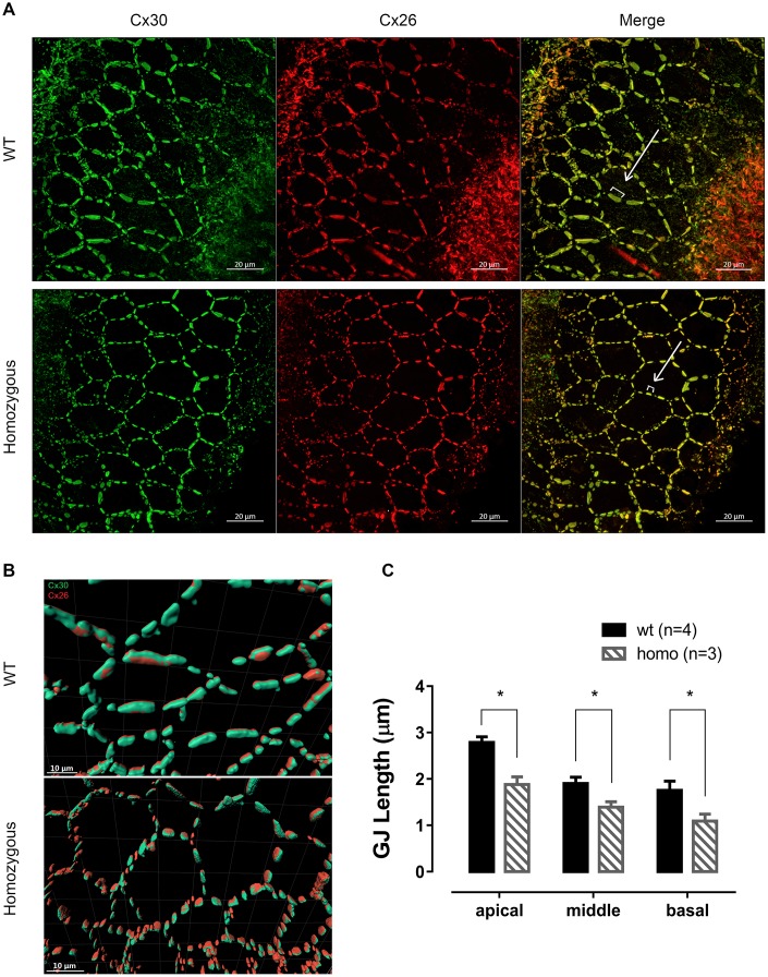
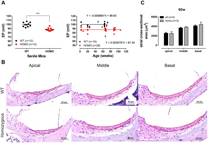
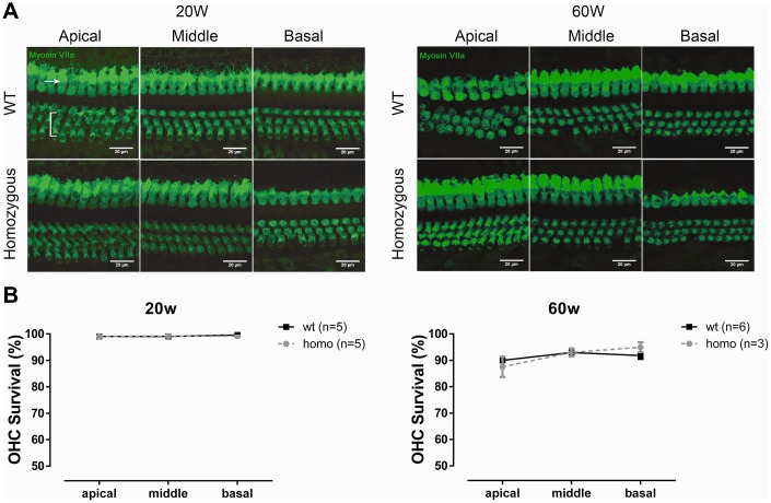
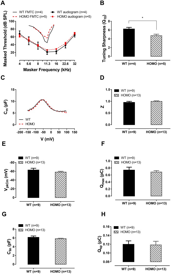
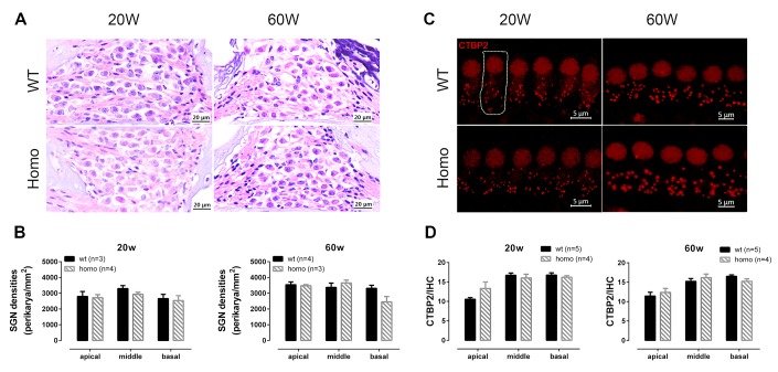
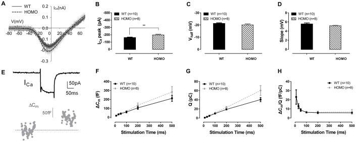
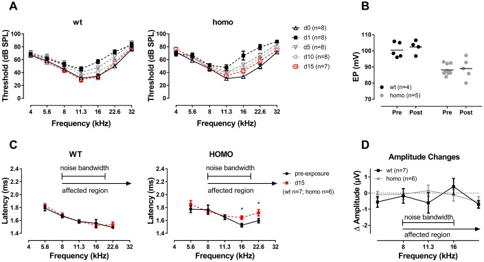
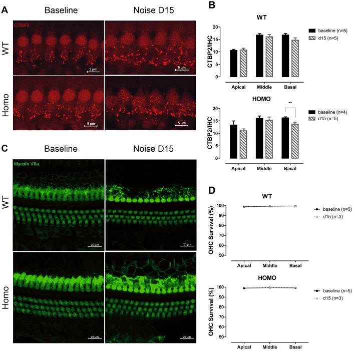
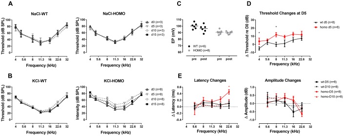
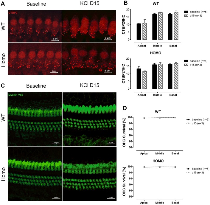
Human p.V37I mutation of gene was strongly correlated with late-onset progressive hearing loss, especially among East Asia populations. We generated a knock-in mouse model based on human p.V37I variant (c.109G>A) that recapitulated the human phenotype. Cochlear pathology revealed no significant hair cell loss, stria vascularis atrophy or spiral ganglion neuron loss, but a significant change in the length of gap junction plaques, which may have contributed to the observed mild endocochlear potential (EP) drop in homozygous mice lasting lifetime. The cochlear amplification in homozygous mice was compromised, but outer hair cells' function remained unchanged, indicating that the reduced amplification was EP- rather than prestin-generated. In addition to ABR threshold elevation, ABR wave I latencies were also prolonged in aged homozygous animals. We found in homozygous IHCs a significant increase in I but no change in Ca efficiency in triggering exocytosis. Environmental insults such as noise exposure, middle ear injection of KCl solution and systemic application of furosemide all exacerbated the pathological phenotype in homozygous mice. We conclude that this mutation-induced hearing loss results from 1) reduced cochlear amplifier caused by lowered EP, 2) IHCs excitotoxicity associated with potassium accumulation around hair cells, and 3) progression induced by environmental insults.
人类基因的p.V37I突变与迟发性进行性听力损失密切相关,尤其是在东亚人群中。我们基于人类p.V37I变体(c.109G>A)构建了一个基因敲入小鼠模型,该模型重现了人类表型。耳蜗病理学检查显示,毛细胞无明显丢失,血管纹无萎缩,螺旋神经节神经元无丢失,但缝隙连接斑的长度有显著变化,这可能是导致纯合小鼠终生观察到的轻度内淋巴电位(EP)下降的原因。纯合小鼠的耳蜗放大功能受损,但外毛细胞的功能保持不变,这表明放大功能降低是由EP而非prestin引起的。除了ABR阈值升高外,老年纯合动物的ABR波I潜伏期也延长。我们发现纯合内毛细胞中I显著增加,但触发胞吐作用的钙效率没有变化。噪声暴露、中耳注射氯化钾溶液和全身应用速尿等环境损伤均加剧了纯合小鼠的病理表型。我们得出结论,这种突变诱导的听力损失是由以下原因导致的:1)EP降低导致耳蜗放大器功能降低;2)内毛细胞兴奋性毒性与毛细胞周围钾离子积累有关;3)环境损伤诱导病情进展。