Chu Jun, Liu Chen-Xu, Song Rui, Li Qing-Lin
Xin'an Key Laboratory of Medicine, Ministry of Education, Anhui University of Chinese Medicine, Hefei, Anhui Province, China.
Neural Regen Res. 2020 Mar;15(3):528-536. doi: 10.4103/1673-5374.266060.
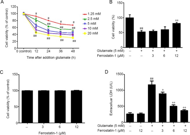
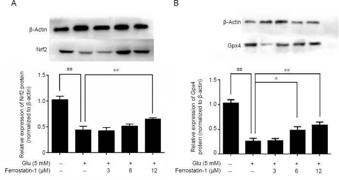
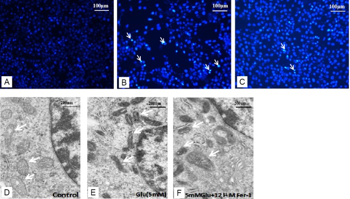
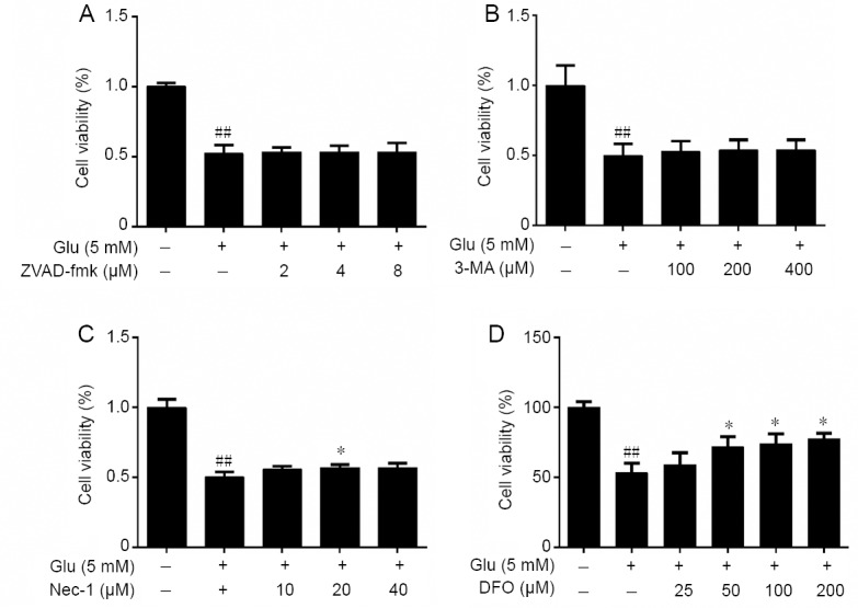
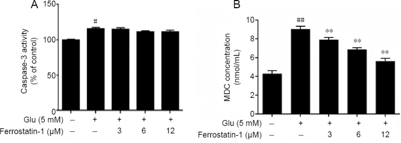
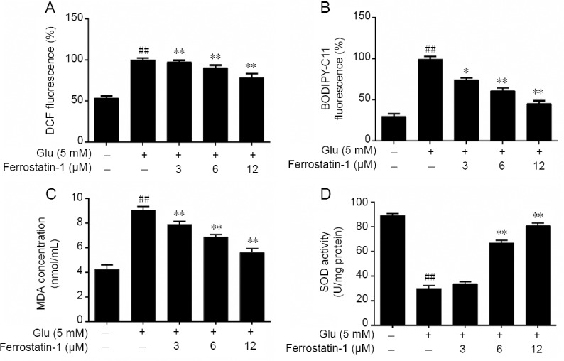
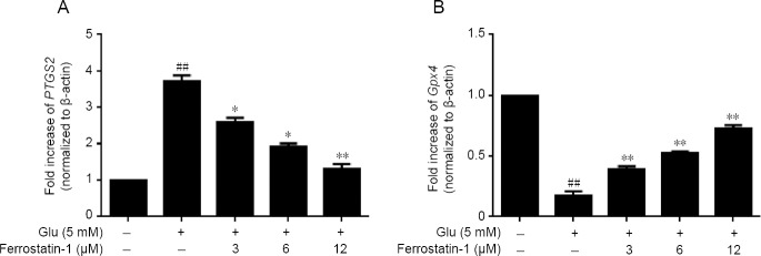
Ferroptosis is a type of programmed cell death dependent on iron. It is different from other forms of cell death such as apoptosis, classic necrosis and autophagy. Ferroptosis is involved in many neurodegenerative diseases. The role of ferroptosis in glutamate-induced neuronal toxicity is not fully understood. To test its toxicity, glutamate (1.25-20 mM) was applied to HT-22 cells for 12 to 48 hours. The optimal experimental conditions occurred at 12 hours after incubation with 5 mM glutamate. Cells were cultured with 3-12 μM ferrostatin-1, an inhibitor of ferroptosis, for 12 hours before exposure to glutamate. The cell viability was detected by 3-(4,5-dimethylthiazol-2-yl)-2,5-diphenyltetrazolium bromide assay. Autophagy was determined by monodansylcadaverine staining and apoptosis by caspase 3 activity. Damage to cell structures was observed under light and by transmission electron microscopy. The release of lactate dehydrogenase was detected by the commercial kit. Reactive oxygen species were measured by flow cytometry. Glutathione peroxidase activity, superoxide dismutase activity and malondialdehyde level were detected by the appropriate commercial kit. Prostaglandin peroxidase synthase 2 and glutathione peroxidase 4 gene expression was detected by real-time quantitative polymerase chain reaction. Glutathione peroxidase 4 and nuclear factor erythroid-derived-like 2 protein expression was detected by western blot analysis. Results showed that ferrostatin-1 can significantly counter the effects of glutamate on HT-22 cells, improving the survival rate, reducing the release of lactate dehydrogenase and reducing the damage to mitochondrial ultrastructure. However, it did not affect the caspase-3 expression and monodansylcadaverine-positive staining in glutamate-injured HT-22 cells. Ferrostatin-1 reduced the levels of reactive oxygen species and malondialdehyde and enhanced superoxide dismutase activity. It decreased gene expression of prostaglandin peroxidase synthase 2 and increased gene expression of glutathione peroxidase 4 and protein expressions of glutathione peroxidase 4 and nuclear factor (erythroid-derived)-like 2 in glutamate-injured HT-22 cells. Treatment of cultured cells with the apoptosis inhibitor Z-Val-Ala-Asp (OMe)-fluoromethyl ketone (2-8 μM), autophagy inhibitor 3-methyladenine (100-400 μM) or necrosis inhibitor necrostatin-1 (10-40 μM) had no effect on glutamate induced cell damage. However, the iron chelator deferoxamine mesylate salt inhibited glutamate induced cell death. Thus, the results suggested that ferroptosis is caused by glutamate-induced toxicity and that ferrostatin-1 protects HT-22 cells from glutamate-induced oxidative toxicity by inhibiting the oxidative stress.
铁死亡是一种依赖铁的程序性细胞死亡。它不同于其他形式的细胞死亡,如凋亡、经典坏死和自噬。铁死亡与许多神经退行性疾病有关。铁死亡在谷氨酸诱导的神经元毒性中的作用尚未完全明确。为了测试其毒性,将谷氨酸(1.25 - 20 mM)作用于HT - 22细胞12至48小时。最佳实验条件是在5 mM谷氨酸孵育12小时后出现。在暴露于谷氨酸之前,将细胞与3 - 12 μM铁死亡抑制剂铁抑素 - 1培养12小时。通过3 -(4,5 - 二甲基噻唑 - 2 - 基)- 2,5 - 二苯基四氮唑溴盐法检测细胞活力。通过单丹磺酰尸胺染色测定自噬,通过半胱天冬酶3活性测定凋亡。在光镜和透射电子显微镜下观察细胞结构损伤。通过商业试剂盒检测乳酸脱氢酶的释放。通过流式细胞术测量活性氧。通过适当的商业试剂盒检测谷胱甘肽过氧化物酶活性、超氧化物歧化酶活性和丙二醛水平。通过实时定量聚合酶链反应检测前列腺素过氧化物酶合酶2和谷胱甘肽过氧化物酶4基因表达。通过蛋白质印迹分析检测谷胱甘肽过氧化物酶4和核因子红细胞衍生样2蛋白表达。结果表明,铁抑素 - 1可以显著对抗谷氨酸对HT - 22细胞的影响,提高存活率,减少乳酸脱氢酶的释放并减少线粒体超微结构损伤。然而,它不影响谷氨酸损伤的HT - 22细胞中半胱天冬酶 - 3的表达和单丹磺酰尸胺阳性染色。铁抑素 - 1降低了活性氧和丙二醛水平,并增强了超氧化物歧化酶活性。它降低了谷氨酸损伤的HT - 22细胞中前列腺素过氧化物酶合酶2的基因表达,并增加了谷胱甘肽过氧化物酶4的基因表达以及谷胱甘肽过氧化物酶4和核因子(红细胞衍生)样2的蛋白表达。用凋亡抑制剂Z - Val - Ala - Asp(OMe)-氟甲基酮(2 - 8 μM)、自噬抑制剂3 - 甲基腺嘌呤(100 - 400 μM)或坏死抑制剂坏死素 - 1(10 - 40 μM)处理培养细胞对谷氨酸诱导的细胞损伤没有影响。然而,铁螯合剂甲磺酸去铁胺抑制了谷氨酸诱导的细胞死亡。因此,结果表明铁死亡是由谷氨酸诱导的毒性引起的,并且铁抑素 - 1通过抑制氧化应激保护HT - 22细胞免受谷氨酸诱导的氧化毒性。