Division of Nephrology and Hypertension, Department of Internal Medicine, The Jikei University School of Medicine, Tokyo, Japan.
Clinical Research Support Center, The Jikei University School of Medicine Tokyo, Tokyo, Japan.
Sci Rep. 2019 Oct 7;9(1):14400. doi: 10.1038/s41598-019-50529-x.
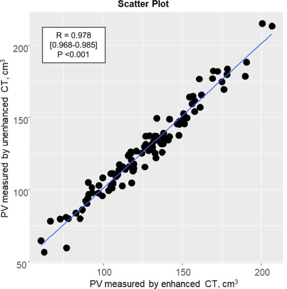
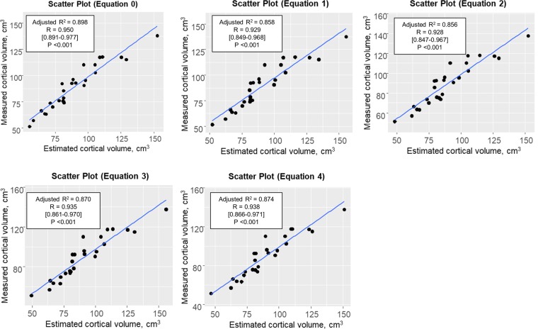
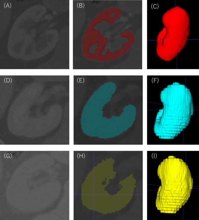
Methods for estimating nephron number in a clinical setting may be useful for predicting renal outcomes. This study aimed to establish such a method using unenhanced computed tomography (CT) and biopsy-based stereology. Patients or living kidney donors simultaneously subjected to enhanced and unenhanced CT examinations were randomly assigned to development and validation groups. The enhanced CT-measured arterial phase and the venous phase images of kidneys were regarded as the true values for cortical volume and parenchymal volume, respectively. Linear multiple regression analysis was used to create models for estimating cortical volume using explanatory variables including unenhanced CT-measured parenchymal volume. Nephron number was determined as the product of cortical volume and the glomerular density in biopsies of donors. Five equations for estimating cortical volume were created and verified. In donors, estimated nephron number by unenhanced CT was consistent with that by enhanced CT, with minimal errors in all models (636-655 ± 210-219 vs. 648 ± 224 × 10/kidney). Clinical characteristics combined with parenchymal volume did not improve the equation over parenchymal volume alone. These results support the feasibility of estimating nephron number by a combination of unenhanced CT and biopsy-based stereology, with a possible application for renal disease patients who are often not suitable for contrast media.
方法估计肾单位数量在临床环境中可能有助于预测肾脏的结果。本研究旨在建立一种方法使用非增强 CT 和基于活检体视学。患者或活体供肾者同时接受增强和非增强 CT 检查被随机分配到发展和验证组。增强 CT 测量的动脉期和静脉期图像的肾脏被认为是皮质体积和实质体积的真实值,分别。线性多元回归分析用于创建模型,使用解释变量包括非增强 CT 测量的实质体积来估计皮质体积。肾单位数量被确定为皮质体积和肾小球密度的乘积在供体的活检。创建并验证了五个方程来估计皮质体积。在供体中,非增强 CT 估计的肾单位数量与增强 CT 一致,所有模型的误差最小(636-655 ± 210-219 vs. 648 ± 224 × 10/肾)。临床特征与实质体积结合并不能改善方程比单纯实质体积。这些结果支持使用非增强 CT 和基于活检的体视学相结合来估计肾单位数量的可行性,对于经常不适合使用造影剂的肾病患者可能有应用价值。