Department of Neurosurgery, Inselspital, Bern University Hospital, Bern, Switzerland.
Department of Neurosurgery, Medical Center University of Freiburg, Freiburg, Germany.
PLoS One. 2019 Oct 9;14(10):e0223484. doi: 10.1371/journal.pone.0223484. eCollection 2019.
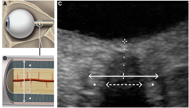
Postural orthostatic tachycardia syndrome is a disorder of the autonomic nervous system. Approximately 30% of patients experience orthostatic headaches. Orthostatic headaches also are a hallmark symptom in spontaneous intracranial hypotension. While the cause of orthostatic headaches in spontaneous intracranial hypotension can be linked to the cerebrospinal fluid loss at the spinal level and consecutively reduced intracranial pressure in the upright position, the cause of orthostatic headaches in postural orthostatic tachycardia syndrome still remains unknown. The present study examined orthostatic changes of intracranial pressure using dynamic ultrasound of the optic nerve and optic nerve sheath diameter in postural orthostatic tachycardia syndrome, spontaneous intracranial hypotension and healthy subjects.
Data was obtained from postural orthostatic tachycardia syndrome patients with (n = 7) and without orthostatic headaches (n = 7), spontaneous intracranial hypotension patients (n = 5) and healthy subjects (n = 8). All participants underwent high-resolution transorbital ultrasound in the supine and upright position to assess optic nerve and optic nerve sheath diameter.
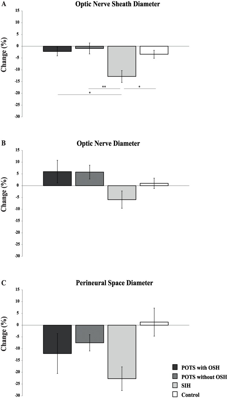
Group differences were found in percentage deviations when changing position of optic nerve sheath diameter (p < 0.01), but not regarding the optic nerve diameter. Pairwise comparisons indicated differences in optic nerve sheath diameter only between spontaneous intracranial hypotension and the other groups. No differences were found between postural orthostatic tachycardia syndrome patients with and without orthostatic headaches.
This study shows that the size of the optic nerve sheath diameter dynamically decreases during orthostatic stress in spontaneous intracranial hypotension, but not in postural orthostatic tachycardia syndrome with or without orthostatic headaches, which indicates different underlying causes.
体位性心动过速综合征是一种自主神经系统疾病。约 30%的患者出现体位性头痛。体位性头痛也是自发性颅内低血压的一个标志性症状。虽然自发性颅内低血压引起的体位性头痛的原因可能与脊柱水平的脑脊液流失以及直立时颅内压力降低有关,但体位性心动过速综合征引起的体位性头痛的原因仍不清楚。本研究使用视神经和视神经鞘直径的动态超声检查,研究体位性心动过速综合征、自发性颅内低血压和健康受试者的颅内压体位变化。
从体位性心动过速综合征伴(n = 7)和不伴体位性头痛(n = 7)、自发性颅内低血压(n = 5)和健康受试者(n = 8)的患者中获取数据。所有参与者均接受高分辨率经眶超声检查,以评估视神经和视神经鞘直径在仰卧位和直立位的变化。
当改变视神经鞘直径的位置时,组间差异存在百分比偏差(p < 0.01),但视神经直径没有差异。成对比较表明,只有自发性颅内低血压与其他组之间存在视神经鞘直径差异。体位性心动过速综合征伴或不伴体位性头痛的患者之间无差异。
本研究表明,在自发性颅内低血压中,视神经鞘直径在直立位时会动态减小,但在体位性心动过速综合征伴或不伴体位性头痛中则不会,这表明其潜在原因不同。