• 文献检索
  • 文档翻译
  • 深度研究
  • 学术资讯
  • Suppr Zotero 插件Zotero 插件
  • 邀请有礼
  • 套餐&价格
  • 历史记录
应用&插件
Suppr Zotero 插件Zotero 插件浏览器插件Mac 客户端Windows 客户端微信小程序
定价
高级版会员购买积分包购买API积分包
服务
文献检索文档翻译深度研究API 文档MCP 服务
关于我们
关于 Suppr公司介绍联系我们用户协议隐私条款
关注我们

Suppr 超能文献

核心技术专利:CN118964589B侵权必究
粤ICP备2023148730 号-1Suppr @ 2026

文献检索

告别复杂PubMed语法,用中文像聊天一样搜索,搜遍4000万医学文献。AI智能推荐,让科研检索更轻松。

立即免费搜索

文件翻译

保留排版,准确专业,支持PDF/Word/PPT等文件格式,支持 12+语言互译。

免费翻译文档

深度研究

AI帮你快速写综述,25分钟生成高质量综述,智能提取关键信息,辅助科研写作。

立即免费体验

利用光学相干断层扫描技术对人心房左室内膜下脂肪组织和纤维方向分布的成像。

Imaging of subendocardial adipose tissue and fiber orientation distributions in the human left atrium using optical coherence tomography.

机构信息

Department of Electrical Engineering, Columbia University, New York, NY.

Department of Pathology, Columbia University Medical Center, New York, NY.

出版信息

J Cardiovasc Electrophysiol. 2019 Dec;30(12):2950-2959. doi: 10.1111/jce.14255. Epub 2019 Nov 5.

DOI:10.1111/jce.14255
PMID:31661178
原文链接:https://pmc.ncbi.nlm.nih.gov/articles/PMC6916589/
Abstract

BACKGROUND

Optical coherence tomography (OCT) has the potential to provide real-time imaging guidance for atrial fibrillation ablation, with promising results for lesion monitoring. OCT can also offer high-resolution imaging of tissue composition, but there is insufficient cardiac OCT data to inform the use of OCT to reveal important tissue architecture of the human left atrium. Thus, the objective of this study was to define OCT imaging data throughout the human left atrium, focusing on the distribution of adipose tissue and fiber orientation as seen from the endocardium.

METHODS AND RESULTS

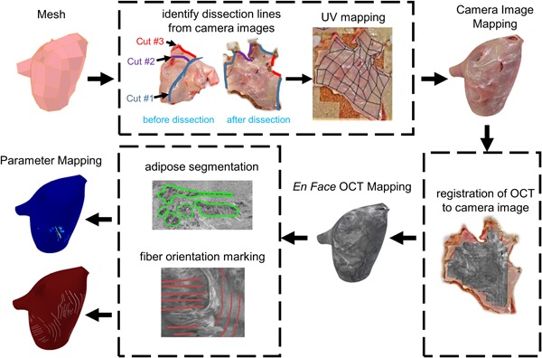
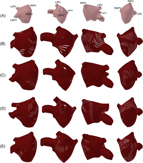
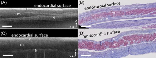
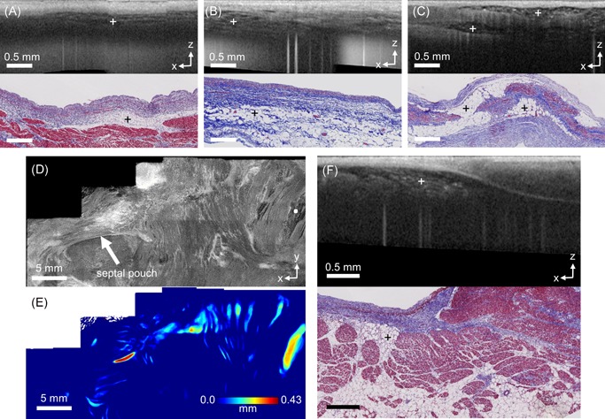
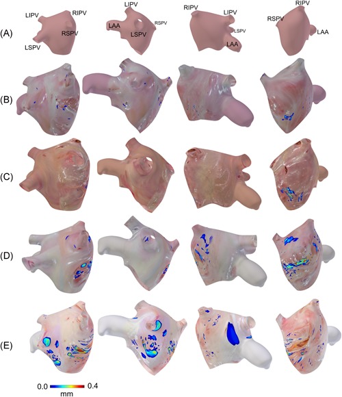
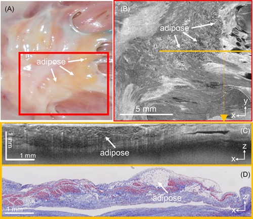
Human hearts (n = 7) were acquired for imaging the left atrium with OCT. A spectral-domain OCT system with 1325 nm center wavelength, 6.5 μm axial resolution, 15 μm lateral resolution, and a maximum imaging depth of 2.51 mm in the air was used. Large-scale OCT image maps of human left atrial tissue were developed, with adipose thickness and fiber orientation extracted from the imaging data. OCT imaging showed scattered distributions of adipose tissue around the septal and pulmonary vein regions, up to a depth of about 0.43 mm from the endocardial surface. The total volume of adipose tissue detected by OCT over one left atrium ranged from 1.42 to 28.74 mm . Limited fiber orientation information primarily around the pulmonary veins and the septum could be identified.

CONCLUSION

OCT imaging could provide adjunctive information on the distribution of subendocardial adipose tissue, particularly around thin areas around the pulmonary veins and septal regions. Variations in OCT-detected tissue composition could potentially assist ablation guidance.

摘要

背景

光学相干断层扫描(OCT)有可能为房颤消融提供实时成像指导,在监测病变方面有很好的效果。OCT 还可以提供组织成分的高分辨率成像,但目前还没有足够的心脏 OCT 数据来告知使用 OCT 揭示人类左心房重要的组织结构。因此,本研究的目的是定义整个左心房的 OCT 成像数据,重点关注从心内膜观察到的脂肪组织分布和纤维方向。

方法和结果

采集人类心脏(n=7)用于左心房 OCT 成像。使用具有 1325nm 中心波长、6.5μm 轴向分辨率、15μm 横向分辨率和在空气中最大成像深度为 2.51mm 的光谱域 OCT 系统。开发了人类左心房组织的大规模 OCT 图像图谱,并从成像数据中提取了脂肪厚度和纤维方向。OCT 成像显示,脂肪组织在间隔和肺静脉区域周围呈散在分布,从心内膜表面深度可达约 0.43mm。通过 OCT 检测到的一个左心房的总脂肪组织体积范围为 1.42 至 28.74mm³。主要在肺静脉和间隔周围可以识别到有限的纤维方向信息。

结论

OCT 成像可以提供心内膜下脂肪组织分布的辅助信息,特别是在肺静脉和间隔区域周围较薄的区域。OCT 检测到的组织成分的变化可能有助于消融指导。

https://cdn.ncbi.nlm.nih.gov/pmc/blobs/595b/6916589/f5498f329338/JCE-30-2950-g006.jpg
https://cdn.ncbi.nlm.nih.gov/pmc/blobs/595b/6916589/1c562ea03220/JCE-30-2950-g001.jpg
https://cdn.ncbi.nlm.nih.gov/pmc/blobs/595b/6916589/7b747966a459/JCE-30-2950-g002.jpg
https://cdn.ncbi.nlm.nih.gov/pmc/blobs/595b/6916589/e3efa09716b0/JCE-30-2950-g003.jpg
https://cdn.ncbi.nlm.nih.gov/pmc/blobs/595b/6916589/035eec16f6fd/JCE-30-2950-g004.jpg
https://cdn.ncbi.nlm.nih.gov/pmc/blobs/595b/6916589/2aa1c41a57a1/JCE-30-2950-g005.jpg
https://cdn.ncbi.nlm.nih.gov/pmc/blobs/595b/6916589/f5498f329338/JCE-30-2950-g006.jpg

相似文献

1
Imaging of subendocardial adipose tissue and fiber orientation distributions in the human left atrium using optical coherence tomography.利用光学相干断层扫描技术对人心房左室内膜下脂肪组织和纤维方向分布的成像。
J Cardiovasc Electrophysiol. 2019 Dec;30(12):2950-2959. doi: 10.1111/jce.14255. Epub 2019 Nov 5.
2
Radiofrequency ablation lesion assessment using optical coherence tomography - a proof-of-concept study.使用光学相干断层扫描评估射频消融损伤——一项概念验证研究。
J Cardiovasc Electrophysiol. 2019 Jun;30(6):934-940. doi: 10.1111/jce.13917. Epub 2019 Mar 29.
3
Endocardial imaging of the left atrium in patients with atrial fibrillation.心房颤动患者左心房的心内膜成像。
J Cardiovasc Electrophysiol. 2004 Feb;15(2):247. doi: 10.1046/j.1540-8167.2004.03335.x.
4
Preferential regional distribution of atrial fibrosis in posterior wall around left inferior pulmonary vein as identified by late gadolinium enhancement cardiac magnetic resonance in patients with atrial fibrillation.心房颤动患者心脏磁共振钆延迟增强显示左肺下静脉后壁的心房纤维化呈区域性优势分布。
Europace. 2018 Dec 1;20(12):1959-1965. doi: 10.1093/europace/euy095.
5
Quantification of fiber orientation in the canine atrial pacemaker complex using optical coherence tomography.应用光学相干断层扫描技术对犬心房起搏复合体中纤维方向进行定量分析。
J Biomed Opt. 2012 Jul;17(7):071309. doi: 10.1117/1.JBO.17.7.071309.
6
Clinical use of AcuNav diagnostic ultrasound catheter imaging during left heart radiofrequency ablation and transcatheter closure procedures.AcuNav诊断超声导管成像在左心射频消融和经导管封堵手术中的临床应用。
J Am Soc Echocardiogr. 2002 Oct;15(10 Pt 2):1301-8. doi: 10.1067/mje.2002.124646.
7
[Value of two left atrium and pulmonary vein stereoscopic imaging reconstruction methods on guiding radiofrequency ablation for atrial fibrillation].两种左心房及肺静脉立体成像重建方法在指导房颤射频消融中的价值
Zhonghua Xin Xue Guan Bing Za Zhi. 2017 Nov 24;45(11):930-934. doi: 10.3760/cma.j.issn.0253-3758.2017.11.007.
8
Tissue-Specific Optical Mapping Models of Swine Atria Informed by Optical Coherence Tomography.基于光相干断层扫描的猪心房组织特异性光学标测模型。
Biophys J. 2018 Mar 27;114(6):1477-1489. doi: 10.1016/j.bpj.2018.01.035.
9
Three-dimensional Integrated Functional, Structural, and Computational Mapping to Define the Structural "Fingerprints" of Heart-Specific Atrial Fibrillation Drivers in Human Heart Ex Vivo.三维综合功能、结构和计算图谱,以确定人心外植体中心脏特异性心房颤动驱动因素的结构“指纹”。
J Am Heart Assoc. 2017 Aug 22;6(8):e005922. doi: 10.1161/JAHA.117.005922.
10
Atrial septal displacement for repair of anomalous pulmonary venous return into the right atrium.房间隔移位用于修复异常肺静脉回流至右心房。
Ann Thorac Surg. 1998 Apr;65(4):1110-4. doi: 10.1016/s0003-4975(97)01419-7.

引用本文的文献

1
Robust, high-density lesion mapping in the left atrium with near-infrared spectroscopy.近红外光谱技术实现左心房高密度、稳健的病灶定位。
J Biomed Opt. 2024 Feb;29(2):028001. doi: 10.1117/1.JBO.29.2.028001. Epub 2024 Feb 28.
2
Cardiac Adipose Tissue Segmentation via Image-Level Annotations.基于图像级标注的心脏脂肪组织分割。
IEEE J Biomed Health Inform. 2023 Jun;27(6):2932-2943. doi: 10.1109/JBHI.2023.3263838. Epub 2023 Jun 6.
3
Cardiac endocardial left atrial substrate and lesion depth mapping using near-infrared spectroscopy.

本文引用的文献

1
Characterization of the human myocardium by optical coherence tomography.光学相干断层成像术对人类心肌的描绘。
J Biophotonics. 2019 Dec;12(12):e201900094. doi: 10.1002/jbio.201900094. Epub 2019 Aug 26.
2
Optical coherence tomography imaging of cardiac substrates.心脏基质的光学相干断层扫描成像
Quant Imaging Med Surg. 2019 May;9(5):882-904. doi: 10.21037/qims.2019.05.09.
3
Integrated RFA/PSOCT catheter for real-time guidance of cardiac radio-frequency ablation.用于心脏射频消融实时引导的集成式射频消融/偏振敏感光学相干断层扫描导管
使用近红外光谱法进行心脏心内膜左心房基质和病变深度测绘。
Biomed Opt Express. 2022 Mar 2;13(4):1801-1819. doi: 10.1364/BOE.451547. eCollection 2022 Apr 1.
4
Quantification of irrigated lesion morphology using near-infrared spectroscopy.使用近红外光谱技术对灌洗后的病灶形态进行定量分析。
Sci Rep. 2021 Oct 11;11(1):20160. doi: 10.1038/s41598-021-99725-8.
5
Anisotropic Mechanical Properties of the Human Uterus Measured by Spherical Indentation.球压入法测量人子宫的各向异性力学性能
Ann Biomed Eng. 2021 Aug;49(8):1923-1942. doi: 10.1007/s10439-021-02769-0. Epub 2021 Apr 20.
Biomed Opt Express. 2018 Nov 20;9(12):6400-6411. doi: 10.1364/BOE.9.006400. eCollection 2018 Dec 1.
4
Radiofrequency ablation lesion assessment using optical coherence tomography - a proof-of-concept study.使用光学相干断层扫描评估射频消融损伤——一项概念验证研究。
J Cardiovasc Electrophysiol. 2019 Jun;30(6):934-940. doi: 10.1111/jce.13917. Epub 2019 Mar 29.
5
Mapping the human pulmonary venoatrial junction with optical coherence tomography.用光学相干断层扫描技术绘制人体肺静脉心房交界处图谱。
Biomed Opt Express. 2019 Jan 7;10(2):434-448. doi: 10.1364/BOE.10.000434. eCollection 2019 Feb 1.
6
Differentiation and quantification of fibrosis, fat and fatty fibrosis in human left atrial myocardium using ex vivo MRI.使用离体 MRI 对人类左心房心肌中的纤维化、脂肪和脂肪性纤维化进行分化和定量。
PLoS One. 2018 Oct 8;13(10):e0205104. doi: 10.1371/journal.pone.0205104. eCollection 2018.
7
Tissue-Specific Optical Mapping Models of Swine Atria Informed by Optical Coherence Tomography.基于光相干断层扫描的猪心房组织特异性光学标测模型。
Biophys J. 2018 Mar 27;114(6):1477-1489. doi: 10.1016/j.bpj.2018.01.035.
8
Serial optical coherence scanning reveals an association between cardiac function and the heart architecture in the aging rodent heart.连续光学相干扫描揭示了衰老啮齿动物心脏中心脏功能与心脏结构之间的关联。
Biomed Opt Express. 2017 Oct 16;8(11):5027-5038. doi: 10.1364/BOE.8.005027. eCollection 2017 Nov 1.
9
Multicontrast endomyocardial imaging by single-channel high-resolution cross-polarization optical coherence tomography.单通道高分辨率交叉偏振光相干断层成像术的多对比心肌内成像。
J Biophotonics. 2018 Apr;11(4):e201700204. doi: 10.1002/jbio.201700204. Epub 2018 Jan 10.
10
Catheterized Fiber-Optics Confocal Microscopy of the Beating Heart In Situ.原位跳动心脏的导管光纤共聚焦显微镜检查。
Circ Cardiovasc Imaging. 2017 Oct;10(10). doi: 10.1161/CIRCIMAGING.117.006881.