Division of Diabetes, Metabolism, and Endocrinology, Department of Medicine, Showa University School of Medicine, Tokyo, 142-8555, Japan.
Department of Anatomy, Showa University School of Medicine, Tokyo, 142-8555, Japan.
Cardiovasc Diabetol. 2019 Oct 31;18(1):143. doi: 10.1186/s12933-019-0947-5.
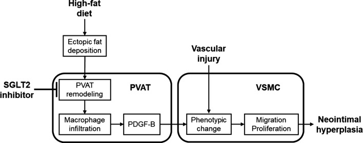
Excess fat deposition could induce phenotypic changes of perivascular adipose tissue (PVAT remodeling), which may promote the progression of atherosclerosis via modulation of adipocytokine secretion. However, it remains unclear whether and how suppression of PVAT remodeling could attenuate vascular injury. In this study, we examined the effect of sodium-glucose cotransporter 2 (SGLT2) inhibitor, luseogliflozin on PVAT remodeling and neointima formation after wire injury in mice.
Wilt-type mice fed with low-fat diet (LFD) or high-fat diet (HFD) received oral administration of luseogliflozin (18 mg/kg/day) or vehicle. Mice underwent bilateral femoral artery wire injury followed by unilateral removal of surrounding PVAT. After 25 days, injured femoral arteries and surrounding PVAT were analyzed.
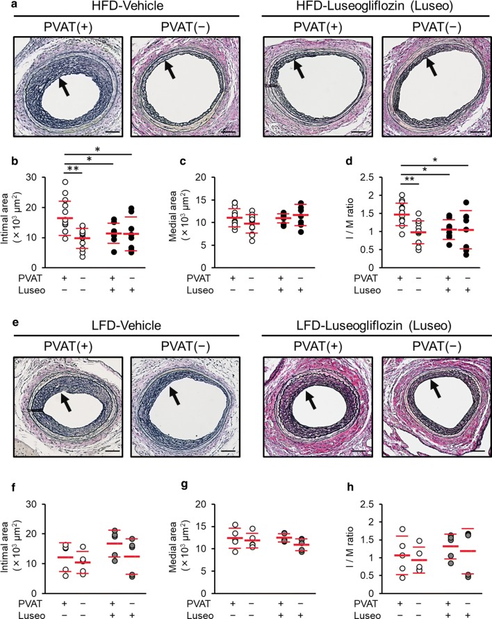
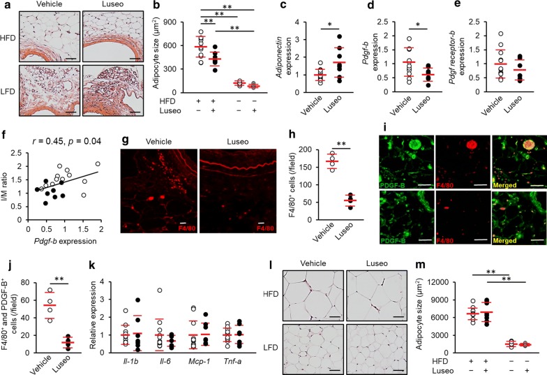
In LFD-fed lean mice, neither luseogliflozin treatment or PVAT removal attenuated the intima-to-media (I/M) ratio of injured arteries. However, in HFD-fed mice, luseogliflozin or PVAT removal reduced the I/M ratio, whereas their combination showed no additive reduction. In PVAT surrounding injured femoral arteries of HFD-fed mice, luseogliflozin treatment decreased the adipocyte sizes. Furthermore, luseogliflozin reduced accumulation of macrophages expressing platelet-derived growth factor-B (PDGF-B) and increased adiponectin gene expression. Gene expression levels of Pdgf-b in PVAT were correlated with the I/M ratio.
Our present study suggests that luseogliflozin could attenuate neointimal hyperplasia after wire injury in HFD-fed mice partly via suppression of macrophage PDGF-B expression in PVAT. Inhibition of PVAT remodeling by luseogliflozin may be a novel therapeutic target for vascular remodeling after angioplasty.
过多的脂肪沉积会引起血管周脂肪组织(PVAT 重塑)的表型改变,这可能通过调节脂肪细胞因子的分泌促进动脉粥样硬化的进展。然而,尚不清楚抑制 PVAT 重塑是否以及如何减轻血管损伤。在这项研究中,我们研究了钠-葡萄糖共转运蛋白 2(SGLT2)抑制剂鲁格列净对高脂饮食喂养的小鼠血管损伤后 PVAT 重塑和新生内膜形成的影响。
低脂肪饮食(LFD)或高脂肪饮食(HFD)喂养的 Wilt 型小鼠接受鲁格列净(18mg/kg/天)或载体的口服给药。小鼠接受双侧股动脉线损伤,随后去除周围的 PVAT。25 天后,分析损伤的股动脉和周围的 PVAT。
在 LFD 喂养的瘦小鼠中,鲁格列净治疗或 PVAT 去除均不能减轻损伤动脉的内膜-中膜(I/M)比值。然而,在 HFD 喂养的小鼠中,鲁格列净或 PVAT 去除降低了 I/M 比值,而两者的组合没有显示出额外的降低。在 HFD 喂养的小鼠损伤股动脉周围的 PVAT 中,鲁格列净治疗降低了脂肪细胞的大小。此外,鲁格列净降低了表达血小板衍生生长因子-B(PDGF-B)的巨噬细胞的积累,并增加了脂联素基因的表达。PVAT 中 Pdgf-b 的基因表达水平与 I/M 比值相关。
本研究表明,鲁格列净可能通过抑制 PVAT 中巨噬细胞 PDGF-B 的表达部分减轻 HFD 喂养的小鼠线损伤后的新生内膜增生。鲁格列净抑制 PVAT 重塑可能是血管成形术后血管重塑的一种新的治疗靶点。