• 文献检索
  • 文档翻译
  • 深度研究
  • 学术资讯
  • Suppr Zotero 插件Zotero 插件
  • 邀请有礼
  • 套餐&价格
  • 历史记录
应用&插件
Suppr Zotero 插件Zotero 插件浏览器插件Mac 客户端Windows 客户端微信小程序
定价
高级版会员购买积分包购买API积分包
服务
文献检索文档翻译深度研究API 文档MCP 服务
关于我们
关于 Suppr公司介绍联系我们用户协议隐私条款
关注我们

Suppr 超能文献

核心技术专利:CN118964589B侵权必究
粤ICP备2023148730 号-1Suppr @ 2026

文献检索

告别复杂PubMed语法,用中文像聊天一样搜索,搜遍4000万医学文献。AI智能推荐,让科研检索更轻松。

立即免费搜索

文件翻译

保留排版,准确专业,支持PDF/Word/PPT等文件格式,支持 12+语言互译。

免费翻译文档

深度研究

AI帮你快速写综述,25分钟生成高质量综述,智能提取关键信息,辅助科研写作。

立即免费体验

成年睾丸中的精原干细胞:特征、调控及潜在应用。

Stem Leydig Cells in the Adult Testis: Characterization, Regulation and Potential Applications.

机构信息

Department of Gynecology and Obstetrics, The Second Affiliated Hospital and Yuying Children's Hospital of Wenzhou Medical University, Wenzhou, Zhejiang, China.

Department of Biochemistry and Molecular Biology, Johns Hopkins Bloomberg School of Public Health, Baltimore, Maryland.

出版信息

Endocr Rev. 2020 Feb 1;41(1):22-32. doi: 10.1210/endrev/bnz013.

DOI:10.1210/endrev/bnz013
PMID:31673697
原文链接:https://pmc.ncbi.nlm.nih.gov/articles/PMC7753054/
Abstract

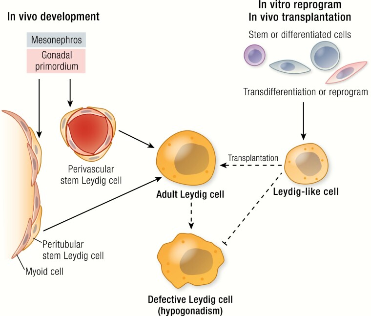
Androgen deficiency (hypogonadism) affects males of all ages. Testosterone replacement therapy (TRT) is effective in restoring serum testosterone and relieving symptoms. TRT, however, is reported to have possible adverse effects in part because administered testosterone is not produced in response to the hypothalamic-pituitary-gonadal (HPG) axis. Progress in stem cell biology offers potential alternatives for treating hypogonadism. Adult Leydig cells (ALCs) are generated by stem Leydig cells (SLCs) during puberty. SLCs persist in the adult testis. Considerable progress has been made in the identification, isolation, expansion and differentiation of SLCs in vitro. In addition to forming ALCs, SLCs are multipotent, with the ability to give rise to all 3 major cell lineages of typical mesenchymal stem cells, including osteoblasts, adipocytes, and chondrocytes. Several regulatory factors, including Desert hedgehog and platelet-derived growth factor, have been reported to play key roles in the proliferation and differentiation of SLCs into the Leydig lineage. In addition, stem cells from several nonsteroidogenic sources, including embryonic stem cells, induced pluripotent stem cells, mature fibroblasts, and mesenchymal stem cells from bone marrow, adipose tissue, and umbilical cord have been transdifferentiated into Leydig-like cells under a variety of induction protocols. ALCs generated from SLCs in vitro, as well as Leydig-like cells, have been successfully transplanted into ALC-depleted animals, restoring serum testosterone levels under HPG control. However, important questions remain, including: How long will the transplanted cells continue to function? Which induction protocol is safest and most effective? For translational purposes, more work is needed with primate cells, especially human.

摘要

雄激素缺乏(性腺功能减退症)影响所有年龄段的男性。睾丸激素替代疗法(TRT)可有效恢复血清睾丸激素并缓解症状。然而,TRT 据报道可能有不良反应,部分原因是给予的睾丸激素不是针对下丘脑-垂体-性腺(HPG)轴产生的。干细胞生物学的进展为治疗性腺功能减退症提供了潜在的替代方法。成年莱迪希细胞(ALC)是由青春期的干细胞莱迪希细胞(SLC)产生的。SLC 存在于成年睾丸中。在体外鉴定、分离、扩增和分化 SLC 方面已经取得了相当大的进展。除了形成 ALC 之外,SLC 还具有多能性,能够产生典型间充质干细胞的所有 3 个主要细胞谱系,包括成骨细胞、脂肪细胞和成软骨细胞。几种调节因子,包括沙漠刺猬和血小板衍生生长因子,已被报道在 SLC 增殖和分化为莱迪希谱系中发挥关键作用。此外,来自几种非甾体来源的干细胞,包括胚胎干细胞、诱导多能干细胞、成熟成纤维细胞以及来自骨髓、脂肪组织和脐带的间充质干细胞,已经在各种诱导方案下转化为莱迪希样细胞。体外培养的 SLC 生成的 ALC 以及莱迪希样细胞已成功移植到 ALC 耗竭的动物体内,在 HPG 控制下恢复血清睾丸激素水平。然而,仍有一些重要问题需要解决,包括:移植细胞能持续多久发挥作用?哪种诱导方案最安全、最有效?为了转化应用,需要使用灵长类细胞,尤其是人类细胞进行更多的工作。

https://cdn.ncbi.nlm.nih.gov/pmc/blobs/4d9b/7753054/b1130730e5ec/bnz013if0001.jpg

相似文献

1
Stem Leydig Cells in the Adult Testis: Characterization, Regulation and Potential Applications.成年睾丸中的精原干细胞:特征、调控及潜在应用。
Endocr Rev. 2020 Feb 1;41(1):22-32. doi: 10.1210/endrev/bnz013.
2
Transplantation of CD51 Stem Leydig Cells: A New Strategy for the Treatment of Testosterone Deficiency.CD51 干细胞睾丸间质细胞移植:治疗睾酮缺乏症的新策略。
Stem Cells. 2017 May;35(5):1222-1232. doi: 10.1002/stem.2569. Epub 2017 Mar 5.
3
Insights into the Regulation on Proliferation and Differentiation of Stem Leydig Cells.深入了解调控精原干细胞增殖与分化的机制
Stem Cell Rev Rep. 2021 Oct;17(5):1521-1533. doi: 10.1007/s12015-021-10133-x. Epub 2021 Feb 17.
4
Endosialin defines human stem Leydig cells with regenerative potential.内唾液酸糖蛋白(endosialin)可鉴定具有再生潜能的人源睾丸间质干细胞。
Hum Reprod. 2020 Oct 1;35(10):2197-2212. doi: 10.1093/humrep/deaa174.
5
Primary human testicular PDGFRα+ cells are multipotent and can be differentiated into cells with Leydig cell characteristics in vitro.原代人睾丸 PDGFRα+ 细胞具有多能性,并且可以在体外分化为具有间质细胞特征的细胞。
Hum Reprod. 2019 Sep 29;34(9):1621-1631. doi: 10.1093/humrep/dez131.
6
Stem Leydig cells: from fetal to aged animals.干细胞样睾丸间质细胞:从胎儿到老年动物
Birth Defects Res C Embryo Today. 2010 Dec;90(4):272-83. doi: 10.1002/bdrc.20192.
7
Characterization of Nestin-positive stem Leydig cells as a potential source for the treatment of testicular Leydig cell dysfunction.巢蛋白阳性的睾丸间质干细胞特性研究:一种治疗睾丸间质细胞功能障碍的潜在细胞来源
Cell Res. 2014 Dec;24(12):1466-85. doi: 10.1038/cr.2014.149. Epub 2014 Nov 21.
8
Leydig cell stem cells: Identification, proliferation and differentiation.睾丸间质干细胞:鉴定、增殖与分化
Mol Cell Endocrinol. 2017 Apr 15;445:65-73. doi: 10.1016/j.mce.2016.10.010. Epub 2016 Oct 12.
9
Restorative functions of Autologous Stem Leydig Cell transplantation in a Testosterone-deficient non-human primate model.自体干细胞莱迪希细胞移植在雄激素缺乏的非人类灵长类动物模型中的修复功能。
Theranostics. 2020 Jul 9;10(19):8705-8720. doi: 10.7150/thno.46854. eCollection 2020.
10
Transplanted human p75-positive stem Leydig cells replace disrupted Leydig cells for testosterone production.移植的人 p75 阳性干细胞莱迪希细胞替代破坏的莱迪希细胞以产生睾酮。
Cell Death Dis. 2017 Oct 12;8(10):e3123. doi: 10.1038/cddis.2017.531.

引用本文的文献

1
Harnessing TGF-β signaling to improve testicular organoid development from dissociated testicular cells.利用转化生长因子-β信号通路改善从解离的睾丸细胞生成睾丸类器官的过程。
Stem Cell Res Ther. 2025 Aug 20;16(1):443. doi: 10.1186/s13287-025-04513-0.
2
Circadian disruption impairs Leydig cell maturation and reproductive development in male rats.昼夜节律紊乱会损害雄性大鼠睾丸间质细胞的成熟和生殖发育。
Reprod Biol Endocrinol. 2025 Jul 17;23(1):104. doi: 10.1186/s12958-025-01440-w.
3
Cre Deleter Mouse Lines for Conditional Knockout of Genes in Testis Cell Types.

本文引用的文献

1
Primary human testicular PDGFRα+ cells are multipotent and can be differentiated into cells with Leydig cell characteristics in vitro.原代人睾丸 PDGFRα+ 细胞具有多能性,并且可以在体外分化为具有间质细胞特征的细胞。
Hum Reprod. 2019 Sep 29;34(9):1621-1631. doi: 10.1093/humrep/dez131.
2
Differentiation of human adipose derived stem cells into Leydig-like cells with molecular compounds.用分子化合物将人脂肪来源干细胞分化为莱迪希样细胞。
J Cell Mol Med. 2019 Sep;23(9):5956-5969. doi: 10.1111/jcmm.14427. Epub 2019 Jul 10.
3
CRISPR/dCas9-mediated activation of multiple endogenous target genes directly converts human foreskin fibroblasts into Leydig-like cells.
用于睾丸细胞类型中基因条件性敲除的Cre删除小鼠品系。
Methods Mol Biol. 2025;2954:287-294. doi: 10.1007/978-1-0716-4698-4_17.
4
Single-cell transcriptomic atlas of the human testis across the reproductive lifespan.人类睾丸在整个生殖寿命期的单细胞转录组图谱
Nat Aging. 2025 Apr;5(4):658-674. doi: 10.1038/s43587-025-00824-2. Epub 2025 Mar 3.
5
Enucleated bone marrow-derived mesenchymal stromal cells regulate immune microenvironment and promote testosterone production through efferocytosis.去核骨髓间充质基质细胞通过胞葬作用调节免疫微环境并促进睾酮生成。
Reprod Biol Endocrinol. 2025 Feb 6;23(1):21. doi: 10.1186/s12958-025-01352-9.
6
The use of deidentified organ donor testes for research.使用经过身份识别去除处理的器官捐献者睾丸进行研究。
Andrology. 2025 Feb 6. doi: 10.1111/andr.70008.
7
Reconstructing the Stem Leydig Cell Niche via the Testicular Extracellular Matrix for the Treatment of Testicular Leydig Cell Dysfunction.通过睾丸细胞外基质重建睾丸间质干细胞微环境以治疗睾丸间质细胞功能障碍
Adv Sci (Weinh). 2025 Jan;12(2):e2410808. doi: 10.1002/advs.202410808. Epub 2024 Nov 18.
8
WNT5A regulates the proliferation, apoptosis and stemness of human stem Leydig cells via the β-catenin signaling pathway.WNT5A 通过β-catenin 信号通路调节人源性精原干细胞的增殖、凋亡和干性。
Cell Mol Life Sci. 2024 Feb 17;81(1):93. doi: 10.1007/s00018-023-05077-z.
9
Morphine compromises androgen biosynthesis by immature Leydig cells from pubertal rat testes in vitro.吗啡在体外损害青春期大鼠睾丸中未成熟睾丸间质细胞的雄激素生物合成。
Toxicol Res (Camb). 2024 Jan 25;13(1):tfae001. doi: 10.1093/toxres/tfae001. eCollection 2024 Feb.
10
The role of HIF-1α-mediated autophagy in ionizing radiation-induced testicular injury.低氧诱导因子 1α 介导体细胞自噬在电离辐射诱导睾丸损伤中的作用。
J Mol Histol. 2023 Oct;54(5):439-451. doi: 10.1007/s10735-023-10153-6. Epub 2023 Sep 20.
CRISPR/dCas9 介导的多个内源性靶基因的激活可直接将人包皮成纤维细胞转化为 Leydig 样细胞。
J Cell Mol Med. 2019 Sep;23(9):6072-6084. doi: 10.1111/jcmm.14470. Epub 2019 Jul 2.
4
Conversion of human fibroblasts into functional Leydig-like cells by small molecules and a single factor.小分子和单一因子将人成纤维细胞转化为功能性的莱迪希样细胞。
Biochem Biophys Res Commun. 2019 Aug 13;516(1):1-7. doi: 10.1016/j.bbrc.2019.05.178. Epub 2019 Jun 8.
5
The Pluripotent Microvascular Pericytes Are the Adult Stem Cells Even in the Testis.多能微血管周细胞是成体干细胞,甚至在睾丸中也是如此。
Adv Exp Med Biol. 2019;1122:235-267. doi: 10.1007/978-3-030-11093-2_13.
6
Differentiation of human induced pluripotent stem cells into Leydig-like cells with molecular compounds.人诱导多能干细胞向莱迪希样细胞的分化与分子化合物。
Cell Death Dis. 2019 Mar 4;10(3):220. doi: 10.1038/s41419-019-1461-0.
7
The Orphan Nuclear Receptors Steroidogenic Factor-1 and Liver Receptor Homolog-1: Structure, Regulation, and Essential Roles in Mammalian Reproduction.孤儿核受体类固醇生成因子-1 和肝受体同源物-1:结构、调控及其在哺乳动物生殖中的重要作用。
Physiol Rev. 2019 Apr 1;99(2):1249-1279. doi: 10.1152/physrev.00019.2018.
8
Luteinizing hormone signaling is involved in synchronization of Leydig cell's clock and is crucial for rhythm robustness of testosterone production†.黄体生成素信号参与了间质细胞时钟的同步,对于睾酮产生的节律稳定性至关重要†。
Biol Reprod. 2019 May 1;100(5):1406-1415. doi: 10.1093/biolre/ioz020.
9
Fetal Leydig cells dedifferentiate and serve as adult Leydig stem cells.胎儿莱迪希细胞去分化并作为成体莱迪希干细胞发挥作用。
Development. 2018 Dec 5;145(23):dev169136. doi: 10.1242/dev.169136.
10
Effects of spermatogenic cycle on Stem Leydig cell proliferation and differentiation.生精周期对精原干细胞增殖和分化的影响。
Mol Cell Endocrinol. 2019 Feb 5;481:35-43. doi: 10.1016/j.mce.2018.11.007. Epub 2018 Nov 23.