Department of Neuroscience, The Scripps Research Institute, Jupiter, FL 33458.
Metabolic Core, The Scripps Research Institute, Jupiter, FL 33458.
Proc Natl Acad Sci U S A. 2019 Nov 19;116(47):23760-23771. doi: 10.1073/pnas.1912868116. Epub 2019 Nov 1.
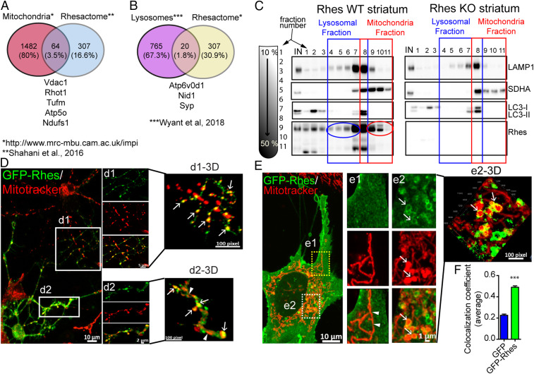
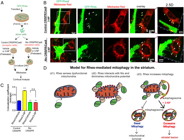
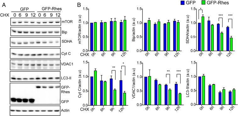
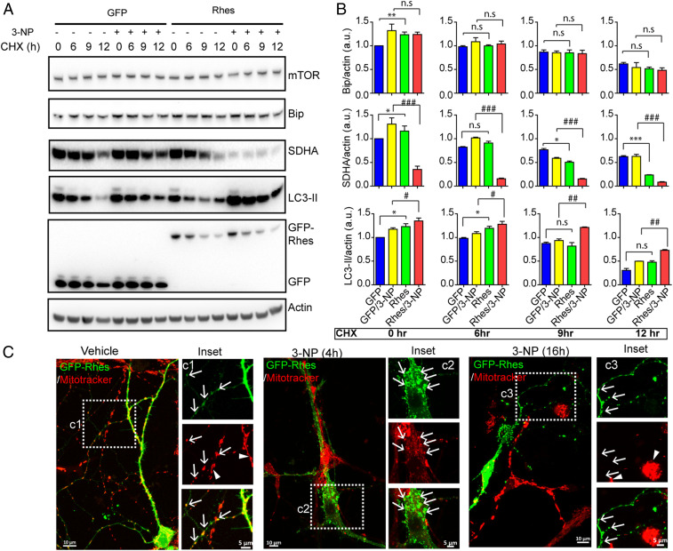
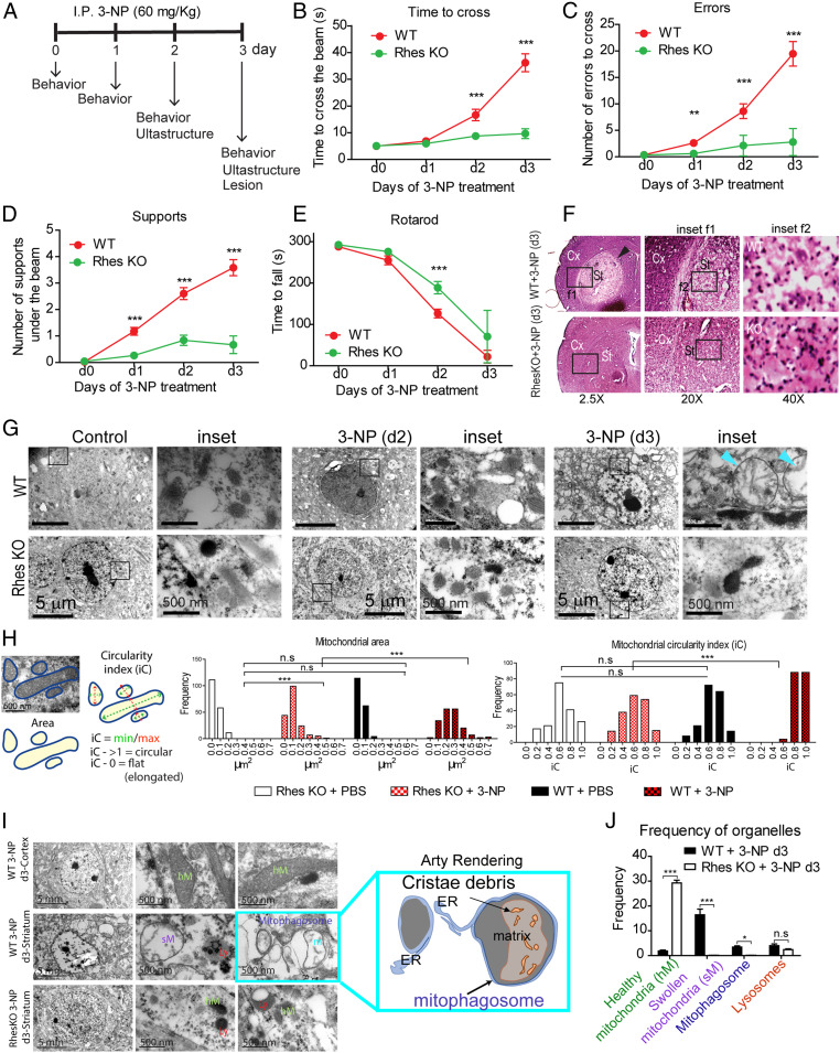
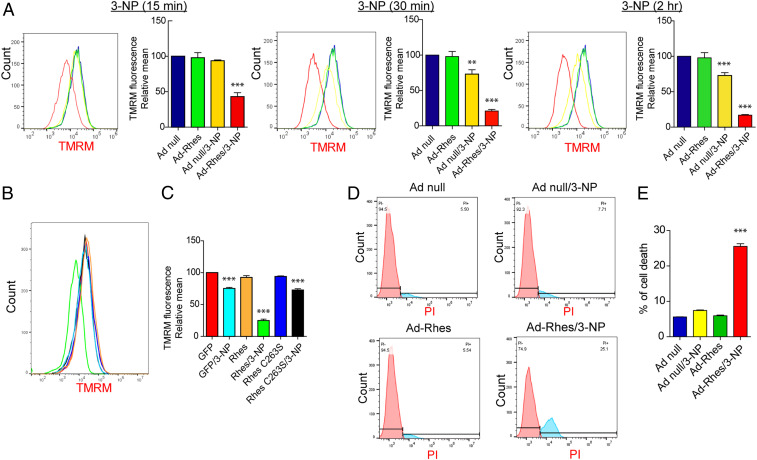
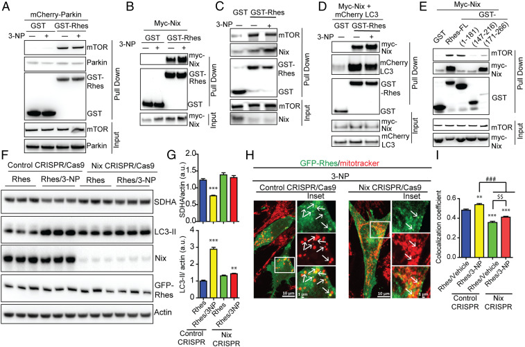
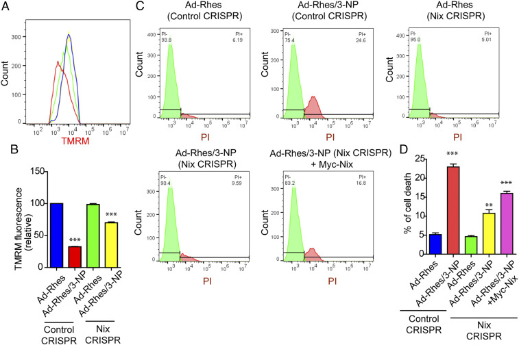
Elimination of dysfunctional mitochondria via mitophagy is essential for cell survival and neuronal functions. But, how impaired mitophagy participates in tissue-specific vulnerability in the brain remains unclear. Here, we find that striatal-enriched protein, Rhes, is a critical regulator of mitophagy and striatal vulnerability in brain. In vivo interactome and density fractionation reveal that Rhes coimmunoprecipitates and cosediments with mitochondrial and lysosomal proteins. Live-cell imaging of cultured striatal neuronal cell line shows Rhes surrounds globular mitochondria, recruits lysosomes, and ultimately degrades mitochondria. In the presence of 3-nitropropionic acid (3-NP), an inhibitor of succinate dehydrogenase, Rhes disrupts mitochondrial membrane potential (ΔΨ ) and promotes excessive mitophagy and cell death. Ultrastructural analysis reveals that systemic injection of 3-NP in mice promotes globular mitochondria, accumulation of mitophagosomes, and striatal lesion only in the wild-type (WT), but not in the Rhes knockout (KO), striatum, suggesting that Rhes is critical for mitophagy and neuronal death in vivo. Mechanistically, Rhes requires Nix (BNIP3L), a known receptor of mitophagy, to disrupt ΔΨ and promote mitophagy and cell death. Rhes interacts with Nix via SUMO E3-ligase domain, and Nix depletion totally abrogates Rhes-mediated mitophagy and cell death in the cultured striatal neuronal cell line. Finally, we find that Rhes, which travels from cell to cell via tunneling nanotube (TNT)-like cellular protrusions, interacts with dysfunctional mitochondria in the neighboring cell in a Nix-dependent manner. Collectively, Rhes is a major regulator of mitophagy via Nix, which may determine striatal vulnerability in the brain.
通过自噬消除功能失调的线粒体对于细胞存活和神经元功能至关重要。但是,受损的自噬如何参与大脑中组织特异性的易损性仍不清楚。在这里,我们发现富含纹状体的蛋白 Rhes 是自噬和大脑纹状体易损性的关键调节因子。体内相互作用组和密度分级分离表明 Rhes 与线粒体和溶酶体蛋白共免疫沉淀和共沉淀。培养的纹状体神经元细胞系的活细胞成像显示 Rhes 包围球状线粒体,招募溶酶体,并最终降解线粒体。在琥珀酸脱氢酶抑制剂 3-硝基丙酸(3-NP)存在的情况下,Rhes 破坏线粒体膜电位(ΔΨ),并促进过度的自噬和细胞死亡。超微结构分析表明,3-NP 在小鼠中的系统注射仅在野生型(WT)而非 Rhes 敲除(KO)纹状体中促进球状线粒体、自噬体的积累和纹状体损伤,表明 Rhes 对于体内自噬和神经元死亡至关重要。在机制上,Rhes 需要 Nix(BNIP3L),一种已知的自噬受体,来破坏 ΔΨ并促进自噬和细胞死亡。Rhes 通过 SUMO E3 连接酶结构域与 Nix 相互作用,并且 Nix 耗竭完全消除了培养的纹状体神经元细胞系中 Rhes 介导的自噬和细胞死亡。最后,我们发现通过隧道纳米管(TNT)样细胞突起在细胞间传递的 Rhes 以 Nix 依赖的方式与相邻细胞中的功能失调线粒体相互作用。总之,Rhes 通过 Nix 是自噬的主要调节剂,这可能决定大脑中纹状体的易损性。