El Hajj Albert, Yacoub Basel, Mansour Mazen, Khauli Raja, Bulbul Mohamad, Nassif Samer, Haidar Mohamad B
Department of Surgery.
Department of Diagnostic Radiology.
Medicine (Baltimore). 2019 Nov;98(44):e17491. doi: 10.1097/MD.0000000000017491.
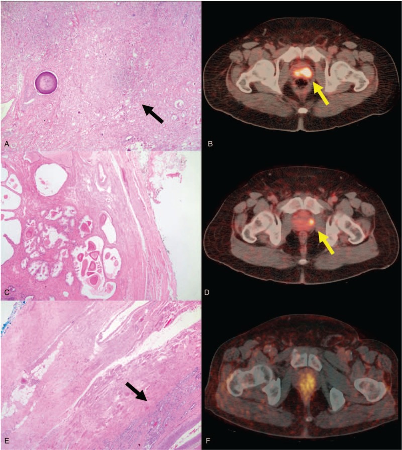
Gallium-68 prostate-specific membrane antigen positron emission tomography-computed tomography (Ga-68 PSMA PET/CT) is an imaging modality that promises improved sensitivity and specificity of detection of prostate cancer lesions based on their increased uptake of PSMA-based radiotracers. It remains an emerging modality that has not yet been endorsed in the guidelines for the management of prostate cancer pending more established evidence to prove its efficacy. The objective of the study is to assess the value of Ga-68 PSMA PET/CT in the detection and localization of patients diagnosed with intermediate or high risk prostate cancer.Twenty three patients with intermediate or high risk prostate cancer had undergone Ga-68 PSMA PET/CT imaging prior to robotic assisted radical prostatectomy. Surgical specimens were then submitted for histological examinations. Lesions visualized on PET/CT and histology were independently mapped unto a 36-segment (Prostate Imaging Reporting and Data System version 2 [PI-RADS v.2]) map of the prostate. Concordance of visualization on PET/CT as compared to the histology as gold standard reference was then assessed. Lesions visualized on PET/CT and histology were independently mapped unto a 36-segment (PI-RADS v.2) map of the prostate. Concordance of visualization on PET/CT as compared to the histology as gold standard reference was then assessed.Sensitivity for all lesions identified on Ga-68 PSMA PET/CT was 42.37%; specificity was 88.61%. Both parameters were higher when considering only index lesions for which sensitivity was 68.42% and specificity was 98.23%. Sensitivity for the index lesions in intermediate risk group was 53.2% and was higher in the high risk group reaching 83.33%.Ga-68 PSMA PET/CT provides accurate localization of tumor lesions in patients with intermediate and high risk prostate cancer.
镓-68前列腺特异性膜抗原正电子发射断层扫描-计算机断层扫描(Ga-68 PSMA PET/CT)是一种成像方式,基于前列腺癌病灶对基于PSMA的放射性示踪剂摄取增加,有望提高其检测的敏感性和特异性。它仍是一种新兴的方式,在有更多确凿证据证明其疗效之前,尚未被纳入前列腺癌管理指南。本研究的目的是评估Ga-68 PSMA PET/CT在诊断为中高危前列腺癌患者的检测和定位中的价值。23例中高危前列腺癌患者在机器人辅助根治性前列腺切除术前行Ga-68 PSMA PET/CT成像。然后将手术标本送检进行组织学检查。将PET/CT和组织学上显示的病灶分别绘制到前列腺的36分区(前列腺影像报告和数据系统第2版[PI-RADS v.2])图谱上。然后以组织学作为金标准参考,评估PET/CT上的显像与组织学的一致性。将PET/CT和组织学上显示的病灶分别绘制到前列腺的36分区(PI-RADS v.2)图谱上。然后以组织学作为金标准参考,评估PET/CT上的显像与组织学的一致性。Ga-68 PSMA PET/CT上识别出的所有病灶的敏感性为42.37%;特异性为88.61%。仅考虑索引病灶时,两个参数均更高,其敏感性为68.42%,特异性为98.23%。中危组索引病灶的敏感性为53.2%,高危组更高,达到83.33%。Ga-68 PSMA PET/CT可为中高危前列腺癌患者的肿瘤病灶提供准确的定位。