Courtney Michael, Johnston Ciaran, Nasoodi Afshin
Department of Radiology, St James's Hospital, Dublin 8, Ireland.
Acta Radiol Open. 2021 Feb 11;10(2):2058460120981001. doi: 10.1177/2058460120981001. eCollection 2021 Feb.
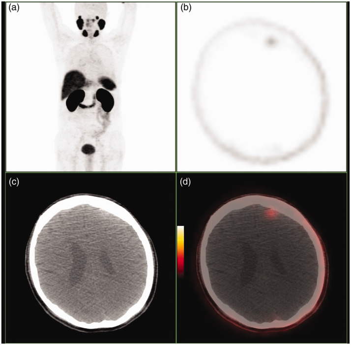
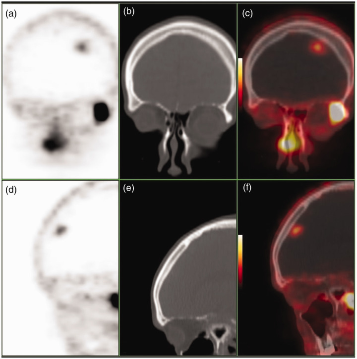
Prostate cancer is the most common malignancy in men with high incidence of recurrence following treatment. Biochemical recurrence, as indicated by rising PSA levels following successful treatment of the primary disease, is a frequent encounter in routine clinical practice. Gallium-PSMA positron emission tomography/computer tomography has been widely accepted as the modality of choice with the highest impact in management of this group of patients. Pitfalls of this diagnostic technique stem from the diversity of histological entities, other than prostate tumour cells, which can demonstrate increased uptake of the radiotracer. We present a case of intracranial uptake of PSMA by meningioma in a patient with BCR, as a pitfall in imaging of prostate cancer. Knowledge of normal distribution of the tracer is of utmost importance when reading positron emission tomography/computer tomography imaging especially given the relative novelty of usage of Gallium-PSMA.
前列腺癌是男性中最常见的恶性肿瘤,治疗后复发率很高。生化复发表现为原发性疾病成功治疗后前列腺特异性抗原(PSA)水平升高,这在常规临床实践中很常见。镓-前列腺特异性膜抗原(PSMA)正电子发射断层扫描/计算机断层扫描已被广泛认为是对这类患者管理影响最大的首选检查方法。这种诊断技术的缺陷源于除前列腺肿瘤细胞外的多种组织学实体,这些实体可表现出放射性示踪剂摄取增加。我们报告一例前列腺癌生化复发患者,其脑膜瘤出现颅内PSMA摄取,这是前列腺癌成像中的一个陷阱。在解读正电子发射断层扫描/计算机断层扫描图像时,了解示踪剂的正常分布至关重要,尤其是考虑到镓-PSMA使用的相对新颖性。