Department of Pediatrics, Division of Hematology and Oncology, University of Nebraska Medical Center, Omaha, NE, 68198, USA.
Department of Biochemistry and Molecular Biology, University of Nebraska Medical Center, Omaha, NE, 68198, USA.
BMC Cancer. 2019 Nov 6;19(1):1056. doi: 10.1186/s12885-019-6291-z.
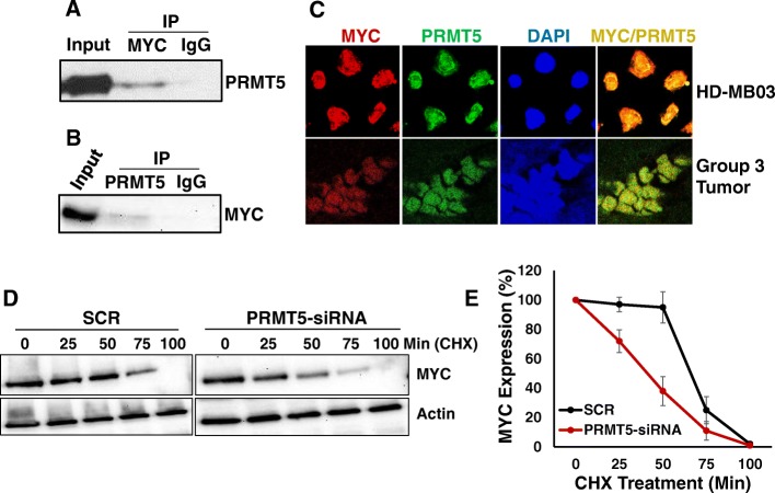
MYC amplification or overexpression is common in Group 3 medulloblastoma and is associated with the worst prognosis. Recently, protein arginine methyl transferase (PRMT) 5 expression has been closely associated with aberrant MYC function in various cancers, including brain tumors such as glioblastoma. However, the role of PRMT5 and its association with MYC in medulloblastoma have not been explored. Here, we report the role of PRMT5 as a novel regulator of MYC and implicate PRMT5 as a potential therapeutic target in MYC-driven medulloblastoma.
Expression and association between PRMT5 and MYC in primary medulloblastoma tumors were investigated using publicly available databases. Expression levels of PRMT5 protein were also examined using medulloblastoma cell lines and primary tumors by western blotting and immunohistochemistry, respectively. Using MYC-driven medulloblastoma cells, we examined the physical interaction between PRMT5 and MYC by co-immunoprecipitation and co-localization experiments. To determine the functional role of PRMT5 in MYC-driven medulloblastoma, PRMT5 was knocked-down in MYC-amplified cells using siRNA and the consequences of knockdown on cell growth and MYC expression/stability were investigated. In vitro therapeutic potential of PRMT5 in medulloblastoma was also evaluated using a small molecule inhibitor, EPZ015666.
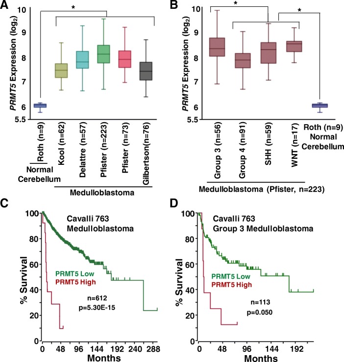
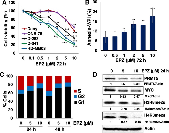
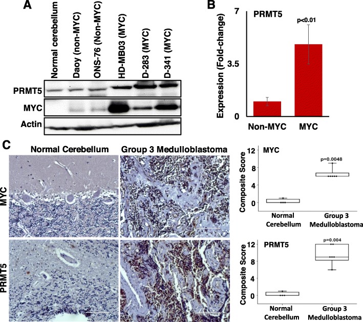
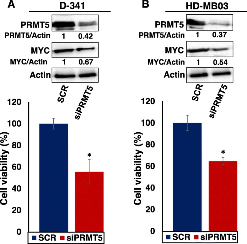
We observed overexpression of PRMT5 in MYC-driven primary medulloblastoma tumors and cell lines compared to non-MYC medulloblastoma tumors and adjacent normal tissues. We also found that high expression of PRMT5 is inversely correlated with patient survival. Knockdown of PRMT5 using siRNA in MYC-driven medulloblastoma cells significantly decreased cell growth and MYC expression. Mechanistically, we found that PRMT5 physically associated with MYC by direct protein-protein interaction. In addition, a cycloheximide chase experiment showed that PRMT5 post-translationally regulated MYC stability. In the context of therapeutics, we observed dose-dependent efficacy of PRMT5 inhibitor EPZ015666 in suppressing cell growth and inducing apoptosis in MYC-driven medulloblastoma cells. Further, the expression levels of PRMT5 and MYC protein were downregulated upon EPZ015666 treatment. We also observed a superior efficacy of this inhibitor against MYC-amplified medulloblastoma cells compared to non-MYC-amplified medulloblastoma cells, indicating specificity.
Our results reveal the regulation of MYC oncoprotein by PRMT5 and suggest that targeting PRMT5 could be a potential therapeutic strategy for MYC-driven medulloblastoma.
MYC 扩增或过表达在 3 组髓母细胞瘤中很常见,与预后最差相关。最近,蛋白质精氨酸甲基转移酶(PRMT)5 的表达与各种癌症中异常 MYC 功能密切相关,包括脑肿瘤如神经胶质瘤。然而,PRMT5 的作用及其与髓母细胞瘤中 MYC 的关联尚未得到探索。在这里,我们报告 PRMT5 作为 MYC 的新型调节因子,并暗示 PRMT5 可能成为 MYC 驱动的髓母细胞瘤的潜在治疗靶点。
使用公开可用的数据库研究 PRMT5 和 MYC 在原发性髓母细胞瘤肿瘤中的表达和关联。通过 Western blot 和免疫组织化学分别在髓母细胞瘤细胞系和原发性肿瘤中检测 PRMT5 蛋白的表达水平。使用 MYC 驱动的髓母细胞瘤细胞,我们通过共免疫沉淀和共定位实验研究 PRMT5 和 MYC 之间的物理相互作用。为了确定 PRMT5 在 MYC 驱动的髓母细胞瘤中的功能作用,使用 siRNA 在 MYC 扩增细胞中敲低 PRMT5,并研究敲低对细胞生长和 MYC 表达/稳定性的影响。还使用小分子抑制剂 EPZ015666 评估 PRMT5 在髓母细胞瘤中的体外治疗潜力。
与非 MYC 髓母细胞瘤肿瘤和相邻正常组织相比,我们观察到 PRMT5 在 MYC 驱动的原发性髓母细胞瘤肿瘤和细胞系中过度表达。我们还发现,PRMT5 的高表达与患者生存呈负相关。在 MYC 驱动的髓母细胞瘤细胞中使用 siRNA 敲低 PRMT5 可显著降低细胞生长和 MYC 表达。在机制上,我们发现 PRMT5 通过直接蛋白质-蛋白质相互作用与 MYC 物理结合。此外,环己酰亚胺追踪实验表明 PRMT5 对 MYC 稳定性进行了翻译后调节。在治疗方面,我们观察到 PRMT5 抑制剂 EPZ015666 以剂量依赖性方式抑制 MYC 驱动的髓母细胞瘤细胞的生长并诱导细胞凋亡。此外,在 EPZ015666 处理后,PRMT5 和 MYC 蛋白的表达水平下调。我们还观察到该抑制剂对 MYC 扩增的髓母细胞瘤细胞的疗效优于非 MYC 扩增的髓母细胞瘤细胞,表明其特异性。
我们的结果揭示了 PRMT5 对 MYC 癌蛋白的调节,并表明靶向 PRMT5 可能是 MYC 驱动的髓母细胞瘤的潜在治疗策略。