Liver Cancer Institute, Zhongshan Hospital, Fudan University & Key Laboratory of Carcinogenesis and Cancer Invasion, Ministry of Education, 136 Yi Xue Yuan Road, Shanghai, 200032, People's Republic of China.
Department of Oncology, Zhongshan Hospital, Fudan University, Shanghai, 200032, People's Republic of China.
J Hematol Oncol. 2019 Nov 8;12(1):112. doi: 10.1186/s13045-019-0795-5.
Increased liver stiffness exerts a detrimental role in driving hepatocellular carcinoma (HCC) malignancy and progression, and indicates a high risk of unfavorable outcomes. However, it remains largely unknown how liver matrix stiffness as an independent cue triggers epithelial-mesenchymal transition (EMT) and facilitates HCC metastasis.
Buffalo rat HCC models with different liver stiffness backgrounds and an in vitro Col I-coated cell culture system with tunable stiffness were used in the study to explore the effects of matrix stiffness on EMT occurrence and its underlying molecular mechanism. Clinical significance of liver stiffness and key molecules required for stiffness-induced EMT were validated in HCC cohorts with different liver stiffness.
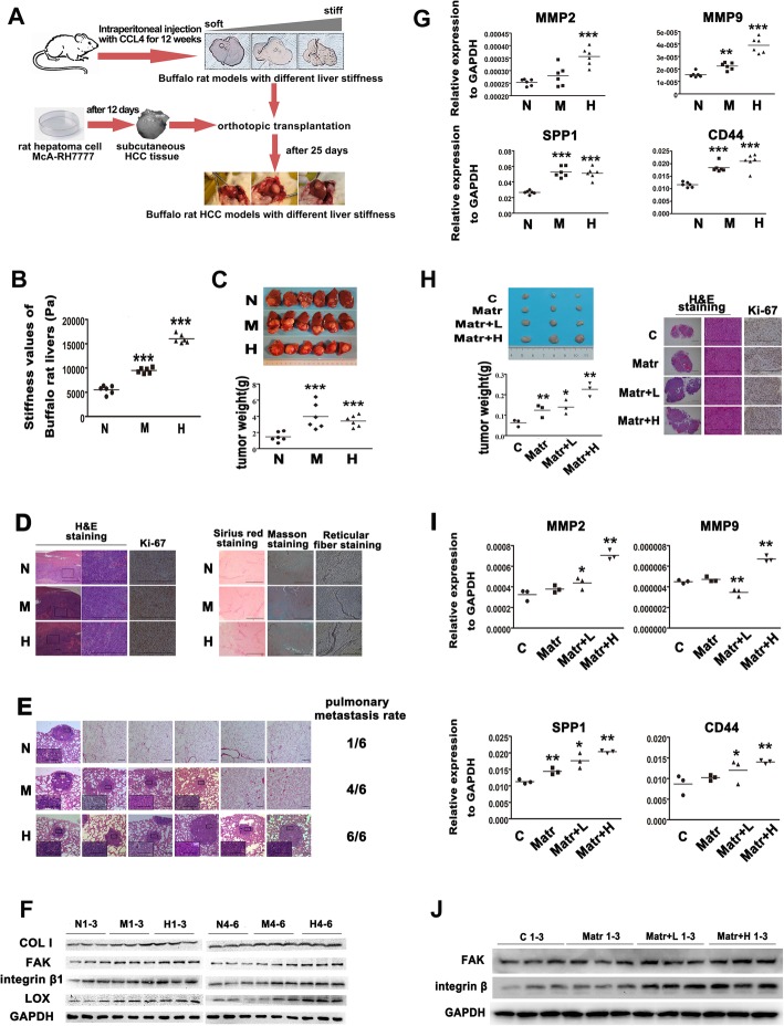
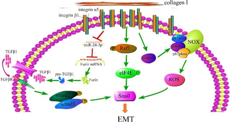
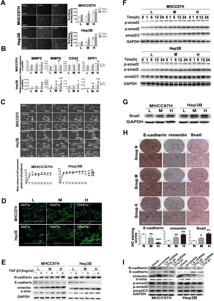
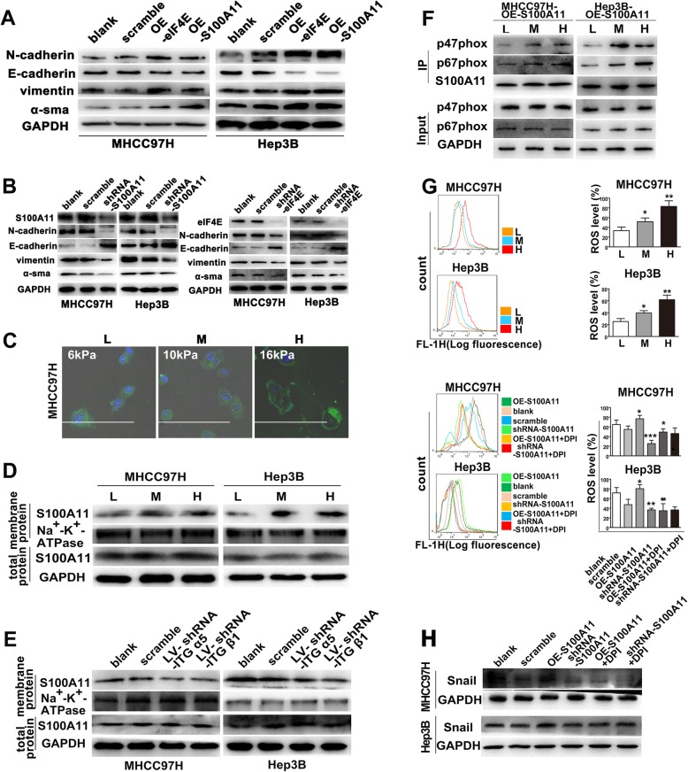
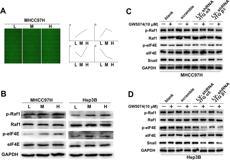
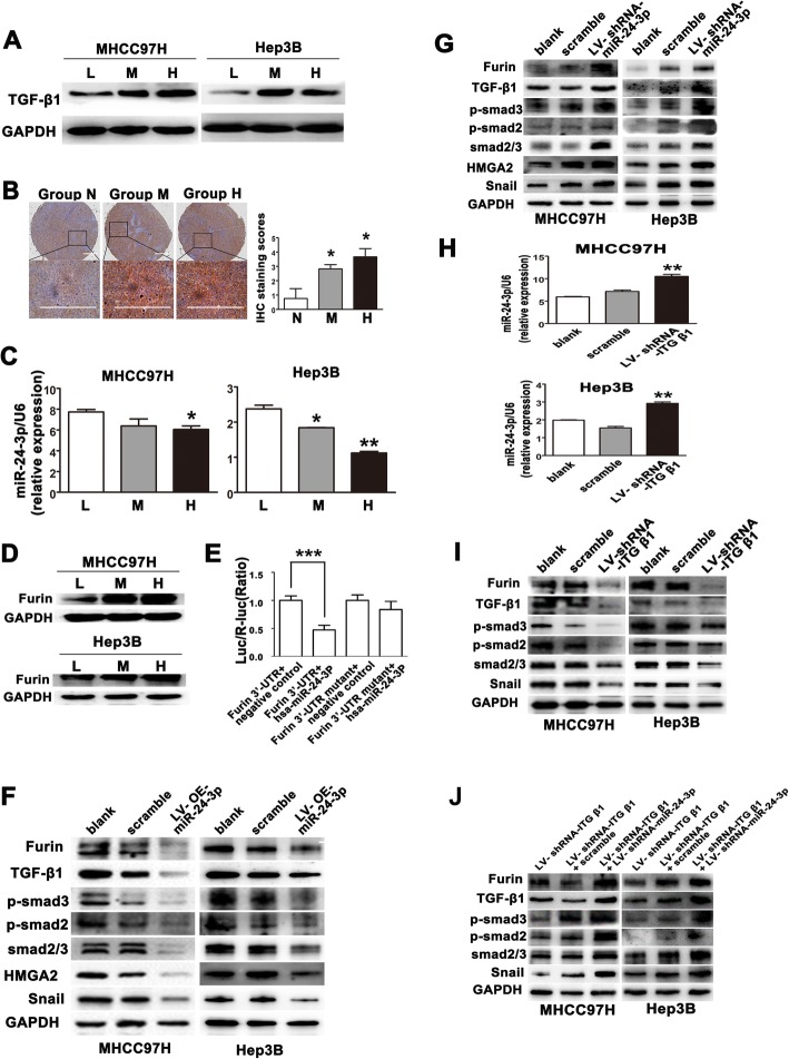
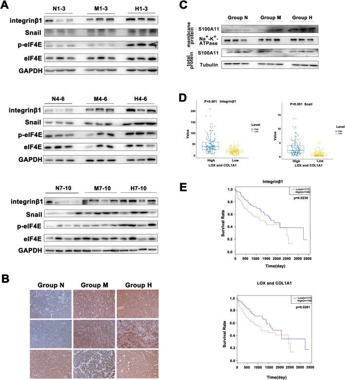
HCC xenografts grown in higher stiffness liver exhibited worse malignant phenotypes and higher lung metastasis rate, suggesting that higher liver stiffness promotes HCC invasion and metastasis. Cell tests in vitro showed that higher matrix stiffness was able to strikingly strengthen malignant phenotypes and independently induce EMT occurrence in HCC cells, and three signaling pathways converging on Snail expression participated in stiffness-mediated effect on EMT including integrin-mediated S100A11 membrane translocation, eIF4E phosphorylation, and TGF β1 autocrine. Additionally, the key molecules required for stiffness-induced EMT were highly expressed in tumor tissues of HCC patients with higher liver stiffness and correlated with poor tumor differentiation and higher recurrence.
Higher matrix stiffness as an initiator triggers epithelial-mesenchymal transition (EMT) in HCC cells independently, and three signaling pathways converging on Snail expression contribute to this pathological process. This work highlights a significant role of biomechanical signal in triggering EMT and facilitating HCC invasion and metastasis.
肝硬度增加对驱动肝细胞癌 (HCC) 恶性和进展具有有害作用,并表明不良结局的风险较高。然而,肝基质硬度作为一个独立的线索如何触发上皮-间充质转化 (EMT) 并促进 HCC 转移,在很大程度上仍不清楚。
本研究使用了具有不同肝硬度背景的水牛大鼠 HCC 模型和具有可调硬度的体外 Col I 涂层细胞培养系统,以探讨基质硬度对 EMT 发生的影响及其潜在的分子机制。在具有不同肝硬度的 HCC 队列中验证了肝硬度和 EMT 所需的关键分子的临床意义。
在肝硬度较高的肝脏中生长的 HCC 异种移植物表现出更差的恶性表型和更高的肺转移率,这表明较高的肝硬度促进了 HCC 的侵袭和转移。体外细胞试验表明,较高的基质硬度能够显著增强恶性表型,并独立诱导 HCC 细胞发生 EMT,参与基质介导 EMT 的三个信号通路包括整合素介导的 S100A11 膜易位、eIF4E 磷酸化和 TGFβ1 自分泌。此外,在肝硬度较高的 HCC 患者的肿瘤组织中,需要用于 EMT 的关键分子高度表达,与肿瘤分化差和复发率高相关。
较高的基质硬度作为启动子独立引发 HCC 细胞的上皮-间充质转化 (EMT),并参与这一病理过程的三个信号通路包括 Snail 表达。这项工作强调了生物力学信号在触发 EMT 和促进 HCC 侵袭和转移中的重要作用。