Centre for Inflammation Research, The Queen's Medical Research Institute, Edinburgh BioQuarter, University of Edinburgh, Edinburgh EH16 4TJ, UK.
Karolinska Institutet (KI), Science for Life Laboratory, Tomtebodavägen 23, Solna 171 65, Sweden.
Cell Rep. 2019 Nov 12;29(7):1832-1847.e8. doi: 10.1016/j.celrep.2019.10.024.
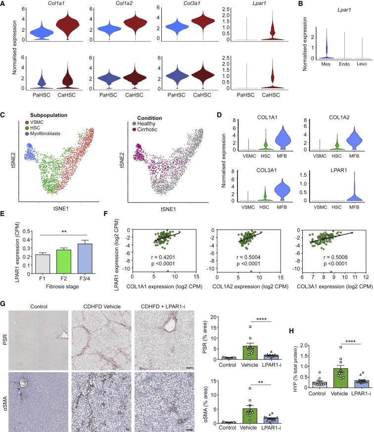
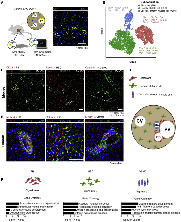
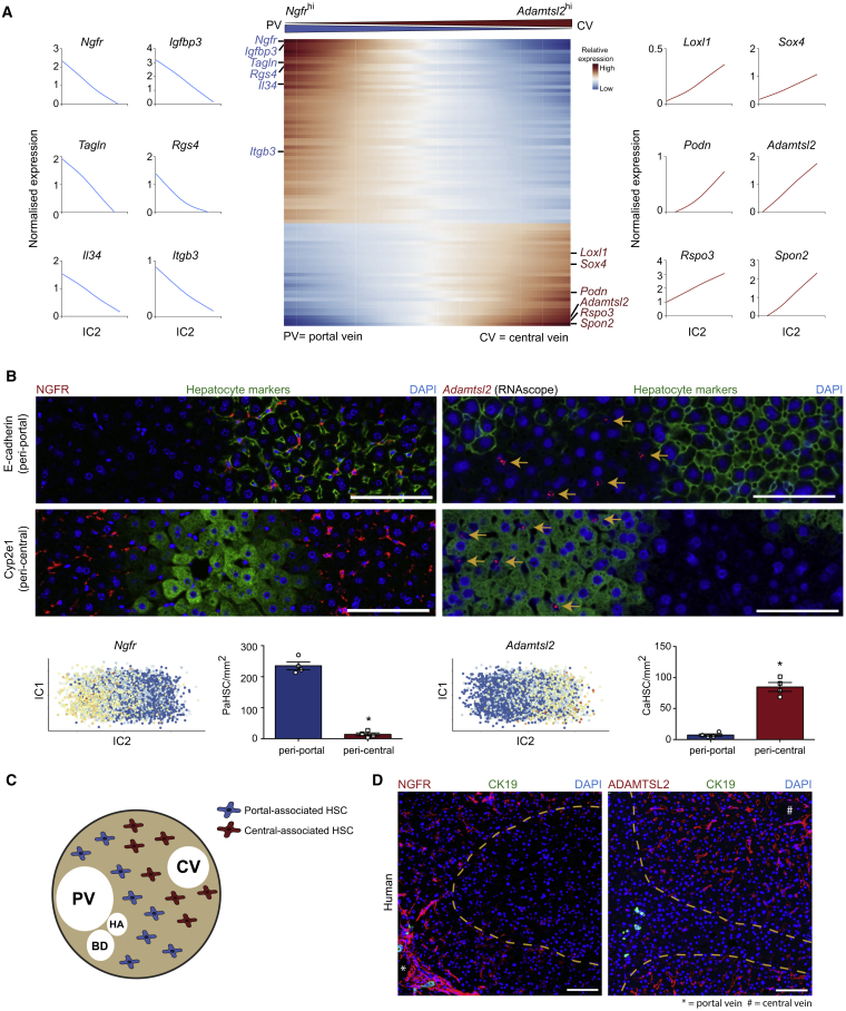
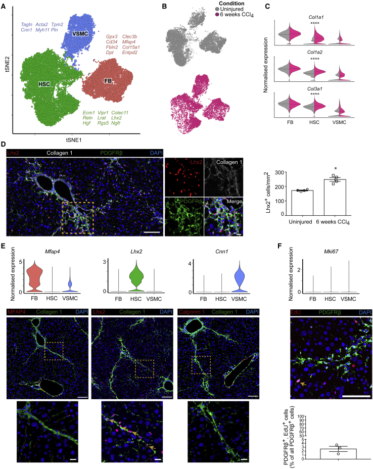
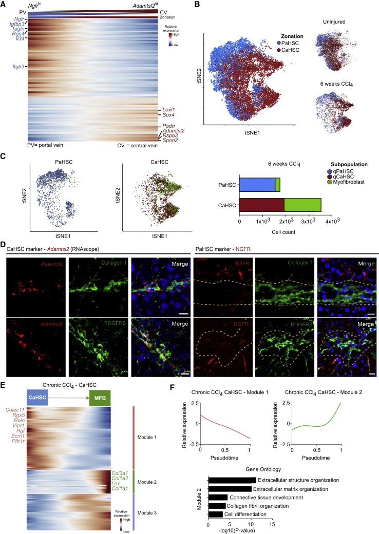
Iterative liver injury results in progressive fibrosis disrupting hepatic architecture, regeneration potential, and liver function. Hepatic stellate cells (HSCs) are a major source of pathological matrix during fibrosis and are thought to be a functionally homogeneous population. Here, we use single-cell RNA sequencing to deconvolve the hepatic mesenchyme in healthy and fibrotic mouse liver, revealing spatial zonation of HSCs across the hepatic lobule. Furthermore, we show that HSCs partition into topographically diametric lobule regions, designated portal vein-associated HSCs (PaHSCs) and central vein-associated HSCs (CaHSCs). Importantly we uncover functional zonation, identifying CaHSCs as the dominant pathogenic collagen-producing cells in a mouse model of centrilobular fibrosis. Finally, we identify LPAR1 as a therapeutic target on collagen-producing CaHSCs, demonstrating that blockade of LPAR1 inhibits liver fibrosis in a rodent NASH model. Taken together, our work illustrates the power of single-cell transcriptomics to resolve the key collagen-producing cells driving liver fibrosis with high precision.
反复的肝损伤导致进行性纤维化,破坏肝结构、再生潜能和肝功能。肝星状细胞(HSCs)是纤维化过程中病理性基质的主要来源,被认为是具有功能均一性的细胞群体。在这里,我们使用单细胞 RNA 测序解析健康和纤维化小鼠肝中的肝间质,揭示 HSCs 在肝小叶内的空间分区。此外,我们还表明 HSCs 分为拓扑上具有直径差异的小叶区域,分别命名为门静脉相关 HSCs(PaHSCs)和中央静脉相关 HSCs(CaHSCs)。重要的是,我们揭示了功能分区,鉴定出 CaHSCs 是小鼠中心小叶纤维化模型中主要的致病变性胶原产生细胞。最后,我们鉴定出 LPAR1 是胶原产生的 CaHSCs 的治疗靶点,证明 LPAR1 阻断可抑制啮齿动物 NASH 模型中的肝纤维化。总之,我们的工作说明了单细胞转录组学在解析驱动肝纤维化的关键胶原产生细胞方面的强大功能,具有高精度。