The Clinical Hospital of Chengdu Brain Science Institute, MOE Key Lab for Neuroinformation, High-Field Magnetic Resonance Brain Imaging Key Laboratory of Sichuan Province, Center for Information in Medicine, School of life Science and technology, University of Electronic Science and Technology of China, Chengdu, 610054, China.
Shanghai Key Laboratory of Psychotic Disorders, Shanghai Mental Health Center, Shanghai Jiaotong University School of Medicine, Shanghai 200030, China.
Neuroimage Clin. 2019;24:102081. doi: 10.1016/j.nicl.2019.102081. Epub 2019 Nov 8.
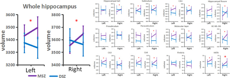
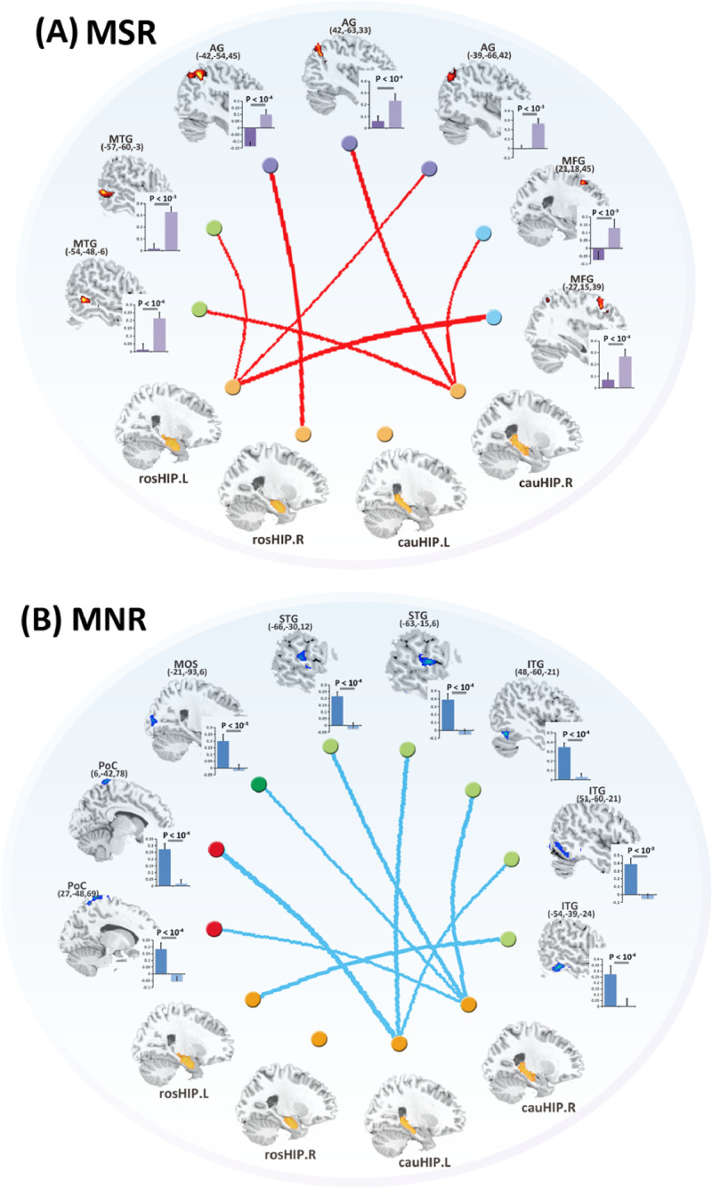
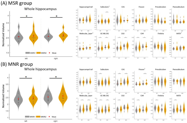
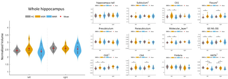
Electroconvulsive therapy (ECT) is considered a treatment option in patients with drug-resistant schizophrenia (SZ). However, approximately one-third of patients do not benefit from ECT in the clinic. Thus, it is critical to investigate differences between ECT responders and non-responders. Accumulated evidence has indicated that one region of ECT action is the hippocampus, which also plays an important role in SZ pathophysiology. To date, no studies have investigated differences in ECT effects in the hippocampus between treatment responders and non-responders. This study recruited twenty-one SZ patients treated for four weeks with ECT (MSZ, n = 21) and twenty-one SZ patients who received pharmaceutical therapy (DSZ, n = 21). The MSZ group was further categorized into responders (MSR, n = 10) or non-responders (MNR, n = 11) based on treatment outcomes by the criterion of a 50% reduction in the Positive and Negative Syndrome Scale total scores. Using structural and resting-state functional MRI, we measured the hippocampal volume and functional connectivity (FC) in all SZ patients (before and after treatment) and 23 healthy controls. In contrast to pharmaceutical therapy, ECT induced bilateral hippocampal volume increases in the MSZ. Both the MSR and MNR exhibited hippocampal expansion after ECT, whereas a lower baseline volume in one of hippocampal subfield (hippocampus-amygdala transition area) was found in the MNR. After ECT, increased FC between the hippocampus and brain networks associated with cognitive function was only observed in the MSR. The mechanism of action of ECT in schizophrenia is complex. A combination of baseline impairment level, ECT-introduced morphological changes and post-ECT FC increases in the hippocampus may jointly contribute to the post-ECT symptom improvements in patients with SZ.
电抽搐治疗(ECT)被认为是治疗耐药性精神分裂症(SZ)患者的一种选择。然而,临床上约有三分之一的患者对 ECT 治疗无效。因此,研究 ECT 应答者和非应答者之间的差异至关重要。大量证据表明,ECT 作用的一个区域是海马体,它在 SZ 病理生理学中也起着重要作用。迄今为止,尚无研究调查 ECT 对海马体的影响在应答者和非应答者之间的差异。本研究招募了 21 名接受 ECT 治疗四周的 SZ 患者(MSZ,n=21)和 21 名接受药物治疗的 SZ 患者(DSZ,n=21)。根据阳性和阴性综合征量表总分降低 50%的标准,MSZ 组进一步分为应答者(MSR,n=10)或无应答者(MNR,n=11)。使用结构和静息态功能磁共振成像,我们测量了所有 SZ 患者(治疗前后)和 23 名健康对照者的海马体体积和功能连接(FC)。与药物治疗不同,ECT 诱导 MSZ 双侧海马体体积增加。MSR 和 MNR 在 ECT 后均表现出海马体扩张,而 MNR 中一个海马亚区(海马-杏仁核过渡区)的基线体积较低。ECT 后,仅在 MSR 中观察到海马体与认知功能相关的脑网络之间的 FC 增加。ECT 在精神分裂症中的作用机制很复杂。基线损伤水平、ECT 引起的形态变化以及 ECT 后海马体 FC 的增加相结合,可能共同导致 SZ 患者 ECT 后的症状改善。