Huaxi MR Research Center (HMRRC), Department of Radiology, West China Hospital of Sichuan University, Chengdu 610041, China.
Huaxi MR Research Center (HMRRC), Department of Radiology, West China Hospital of Sichuan University, Chengdu 610041, China.
Neuroimage Clin. 2018 Jul 10;20:169-176. doi: 10.1016/j.nicl.2018.07.008. eCollection 2018.
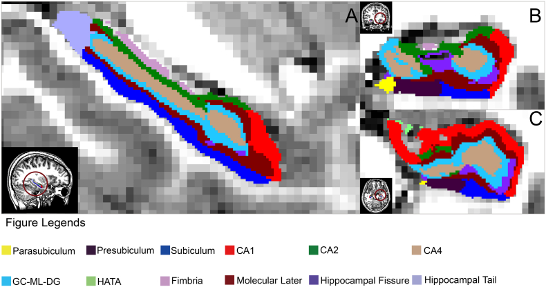
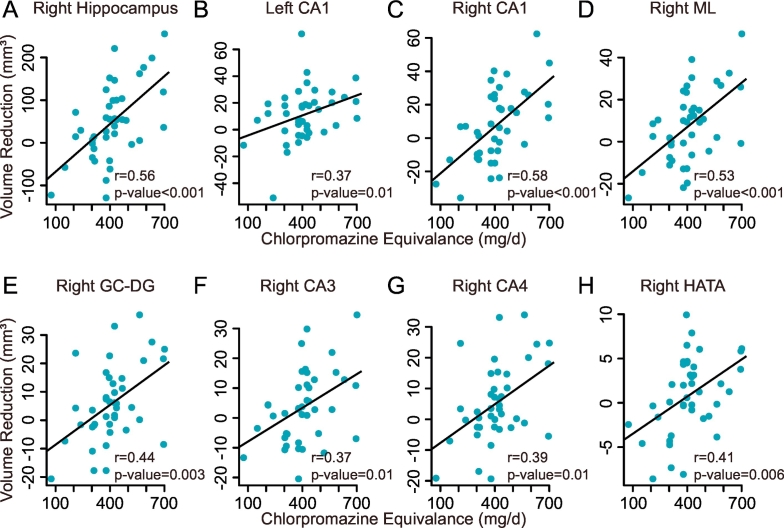
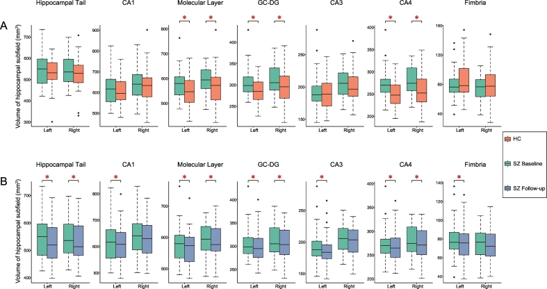
The nature of hippocampal changes in schizophrenia before first treatment, and whether hippocampal subfields are affected by antipsychotic treatment are important questions for schizophrenia research. Forty-one first-episode antipsychotic-naïve acutely ill schizophrenia inpatients had MRI scans before and six weeks after antipsychotic treatment. Thirty-nine matched healthy controls were also scanned, twenty-two of which were scanned a second time six weeks later. Volumes of hippocampal subfields were measured via FreeSurfer v6.0 using a longitudinal analysis pipeline. Before treatment, schizophrenia patients had no significant changes in total hippocampal volume but exhibited significantly greater subfield volumes than controls in bilateral molecular layers of the hippocampus (ML), bilateral granular cell layers of the dentate gyrus (GC-DG), and bilateral cornu ammonis area 4 (CA4). After six weeks of antipsychotic treatment, patients showed volume reductions compared with pretreatment scans in total hippocampus bilaterally, with subfield volume reduction noted in previously enlarged subfields (i.e., bilateral ML, GC-DG and CA4) and in bilateral hippocampal tails, left CA1, CA3, and fimbria. Subfields with volume increases before treatment were reduced to the level of healthy controls (bilateral ML and GC-DG) or near to it (bilateral CA4) after treatment. These results indicate subfield-specific hippocampal hypertrophy prior to treatment, and that these abnormalities were reduced after acute antipsychotic therapy in a dose-related manner together with volume reductions in other areas that were not hypertrophic before treatment.
精神分裂症患者在首次治疗前海马变化的性质,以及海马亚区是否受抗精神病药物治疗的影响,这些都是精神分裂症研究的重要问题。41 名首次接受抗精神病药物治疗的急性精神分裂症住院患者在接受抗精神病药物治疗前和治疗后 6 周进行了 MRI 扫描。还对 39 名匹配的健康对照者进行了扫描,其中 22 名在 6 周后进行了第二次扫描。使用纵向分析管道,通过 FreeSurfer v6.0 测量海马亚区的体积。在治疗前,精神分裂症患者的总海马体积没有明显变化,但双侧海马分子层(ML)、双侧齿状回颗粒细胞层(GC-DG)和双侧角回 4 区(CA4)的亚区体积明显大于对照组。在接受 6 周抗精神病药物治疗后,与治疗前扫描相比,患者双侧总海马体积减小,先前增大的亚区(即双侧 ML、GC-DG 和 CA4)和双侧海马尾部、左侧 CA1、CA3 和穹窿体积减小。治疗前体积增加的亚区在治疗后减小到健康对照组的水平(双侧 ML 和 GC-DG)或接近该水平(双侧 CA4)。这些结果表明,在治疗前存在特定于亚区的海马肥大,并且这些异常在急性抗精神病治疗后以剂量相关的方式减少,同时也减少了治疗前没有肥大的其他区域的体积。