Cell Biology and Biophysics Unit, Porter Neuroscience Research Center, National Institute of Neurological Disorders and Stroke, Bethesda, MD 20982, USA.
Cell Biology and Biophysics Unit, Porter Neuroscience Research Center, National Institute of Neurological Disorders and Stroke, Bethesda, MD 20982, USA; Biochemistry and Biophysics Center, National Heart Lung and Blood Institute, Bethesda, MD 20892, USA.
Dev Cell. 2020 Jan 6;52(1):118-131.e6. doi: 10.1016/j.devcel.2019.10.010. Epub 2019 Nov 14.
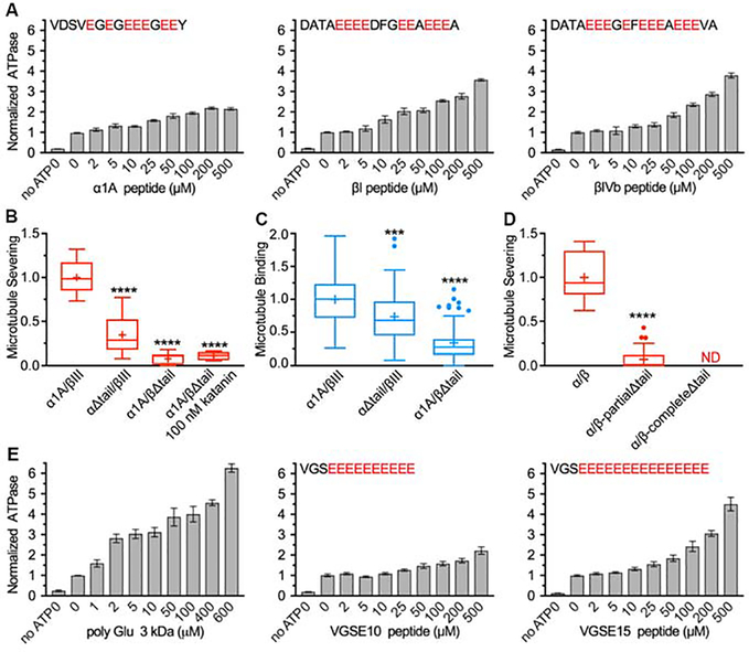
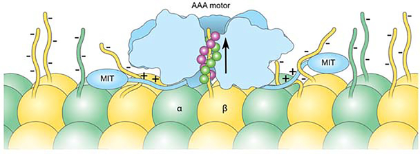
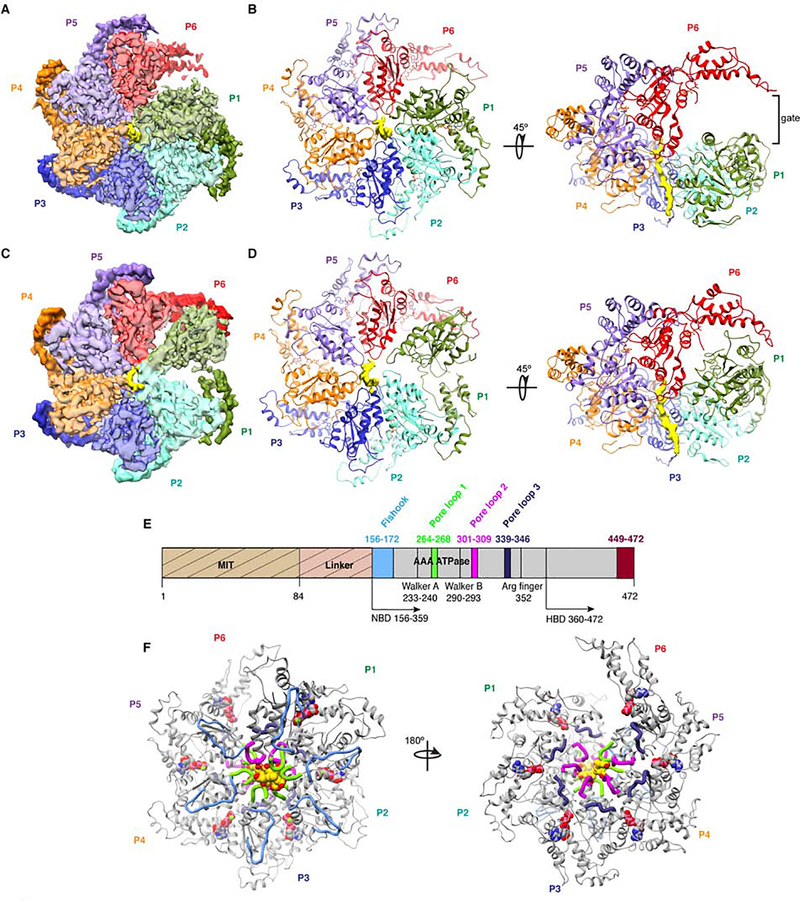
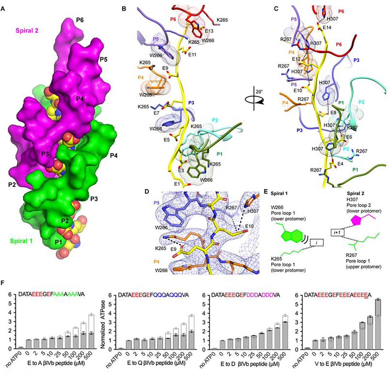
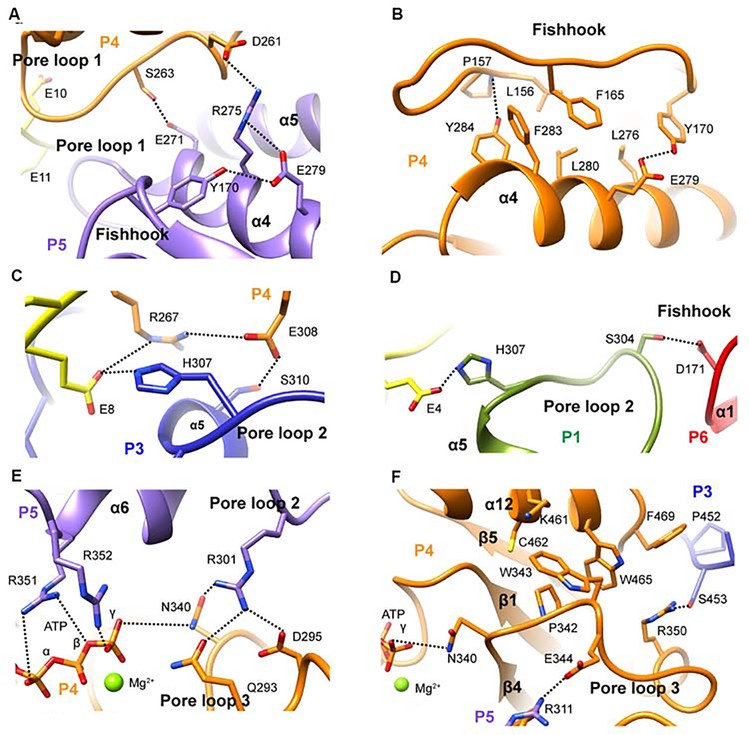
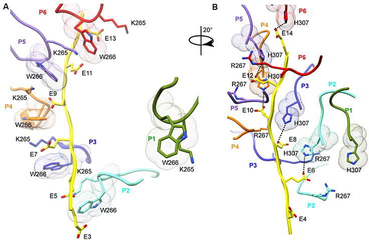
The AAA ATPase katanin severs microtubules. It is critical in cell division, centriole biogenesis, and neuronal morphogenesis. Its mutation causes microcephaly. The microtubule templates katanin hexamerization and activates its ATPase. The structural basis for these activities and how they lead to severing is unknown. Here, we show that β-tubulin tails are necessary and sufficient for severing. Cryoelectron microscopy (cryo-EM) structures reveal the essential tubulin tail glutamates gripped by a double spiral of electropositive loops lining the katanin central pore. Each spiral couples allosterically to the ATPase and binds alternating, successive substrate residues, with consecutive residues coordinated by adjacent protomers. This tightly couples tail binding, hexamerization, and ATPase activation. Hexamer structures in different states suggest an ATPase-driven, ratchet-like translocation of the tubulin tail through the pore. A disordered region outside the AAA core anchors katanin to the microtubule while the AAA motor exerts the forces that extract tubulin dimers and sever the microtubule.
AAA ATP 酶 katánin 可切断微管。它在细胞分裂、中心体发生和神经元形态发生中至关重要。其突变可导致小头畸形。微管模板 katánin 六聚体化并激活其 ATP 酶。这些活性的结构基础以及它们如何导致切断尚不清楚。在这里,我们表明微管尾部是必需的和充分的切断。低温电子显微镜(cryo-EM)结构揭示了必需的微管尾部谷氨酸被包夹在沿着 katánin 中央孔排列的带正电环的双螺旋中。每个螺旋通过变构与 ATP 酶偶联,并结合交替的、连续的底物残基,连续的残基由相邻的亚基协调。这将尾部结合、六聚体化和 ATP 酶激活紧密地偶联起来。不同状态的六聚体结构表明,微管尾部通过孔的 ATP 酶驱动的棘轮样易位。AAA 核心之外的一个无序区域将 katánin 锚定到微管上,而 AAA 马达则施加将微管二聚体提取并切断微管的力。