• 文献检索
  • 文档翻译
  • 深度研究
  • 学术资讯
  • Suppr Zotero 插件Zotero 插件
  • 邀请有礼
  • 套餐&价格
  • 历史记录
应用&插件
Suppr Zotero 插件Zotero 插件浏览器插件Mac 客户端Windows 客户端微信小程序
定价
高级版会员购买积分包购买API积分包
服务
文献检索文档翻译深度研究API 文档MCP 服务
关于我们
关于 Suppr公司介绍联系我们用户协议隐私条款
关注我们

Suppr 超能文献

核心技术专利:CN118964589B侵权必究
粤ICP备2023148730 号-1Suppr @ 2026

文献检索

告别复杂PubMed语法,用中文像聊天一样搜索,搜遍4000万医学文献。AI智能推荐,让科研检索更轻松。

立即免费搜索

文件翻译

保留排版,准确专业,支持PDF/Word/PPT等文件格式,支持 12+语言互译。

免费翻译文档

深度研究

AI帮你快速写综述,25分钟生成高质量综述,智能提取关键信息,辅助科研写作。

立即免费体验

光学偏振检测在牙体硬组织疾病特征分析中的应用。

Optical Polarimetric Detection for Dental Hard Tissue Diseases Characterization.

机构信息

Biomedical Optical Imaging Lab, Department of Photonics, College of Electrical and Computer Engineering, National Chiao Tung University, No.1001, University Road, East District, Hsinchu City 30010, Taiwan.

Department of Stomatology, Taipei Veterans General Hospital, No. 201, Section 2, Shipai Road, Beitou District, Taipei City 11217, Taiwan.

出版信息

Sensors (Basel). 2019 Nov 14;19(22):4971. doi: 10.3390/s19224971.

DOI:10.3390/s19224971
PMID:31739635
原文链接:https://pmc.ncbi.nlm.nih.gov/articles/PMC6891417/
Abstract

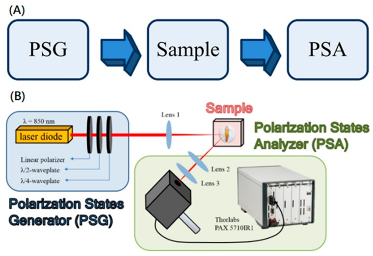
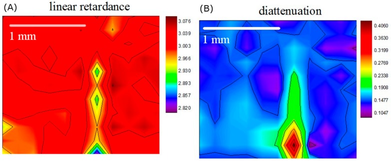
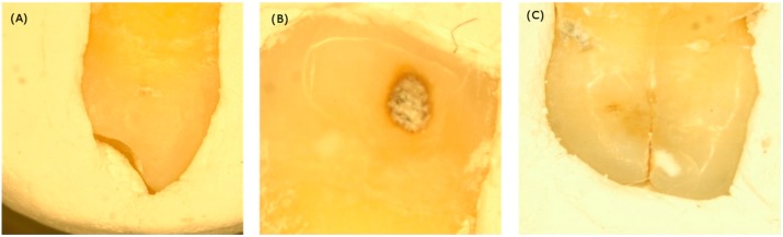
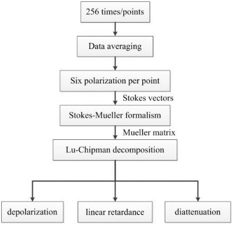
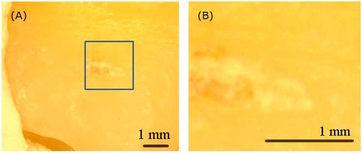
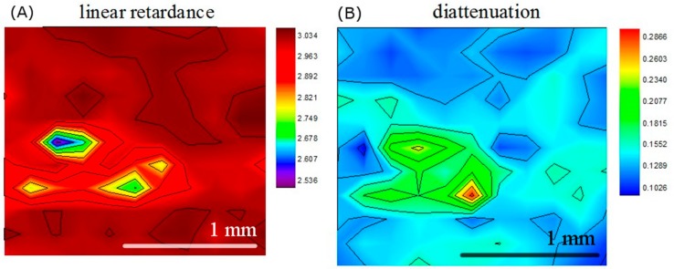
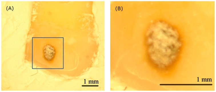
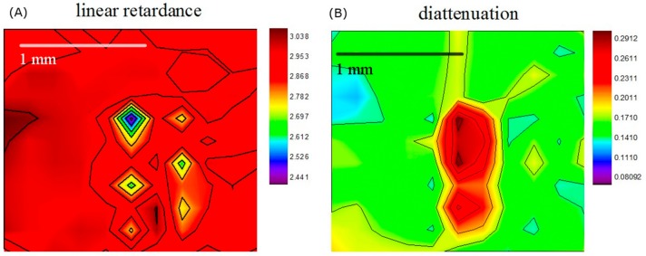
Dental enamel constitutes the outer layer of a crown of teeth and grows nearly parallel. This unique nanostructure makes enamel possess birefringence properties. Currently, there is still no appropriate clinical solution to examine dental hard tissue diseases. Therefore, we developed an optical polarization imaging system for diagnosing dental calculus, caries, and cracked tooth syndrome. By obtaining Stokes signals reflected from samples, Mueller images were constructed and analyzed using Lu-Chipman decomposition. The results showed that diattenuation and linear retardance images can distinguish abnormal tissues. Our result also aligns with previous studies assessed by other methods. Polarimetric imaging is promising for real-time diagnosing.

摘要

牙釉质构成了牙齿冠的外层,并且几乎平行生长。这种独特的纳米结构使牙釉质具有双折射特性。目前,仍然没有合适的临床解决方案来检查牙体硬组织疾病。因此,我们开发了一种用于诊断牙石、龋齿和牙隐裂综合征的光学偏振成像系统。通过获取样品反射的斯托克斯信号,构建穆勒图像,并使用 Lu-Chipman 分解进行分析。结果表明,双折射和线性延迟图像可以区分异常组织。我们的结果与以前使用其他方法评估的研究结果一致。偏振成像有望用于实时诊断。

https://cdn.ncbi.nlm.nih.gov/pmc/blobs/a2f7/6891417/6f2ccd30c689/sensors-19-04971-g009.jpg
https://cdn.ncbi.nlm.nih.gov/pmc/blobs/a2f7/6891417/95f35f458056/sensors-19-04971-g001.jpg
https://cdn.ncbi.nlm.nih.gov/pmc/blobs/a2f7/6891417/73199862ce91/sensors-19-04971-g002.jpg
https://cdn.ncbi.nlm.nih.gov/pmc/blobs/a2f7/6891417/bf52804305fc/sensors-19-04971-g003.jpg
https://cdn.ncbi.nlm.nih.gov/pmc/blobs/a2f7/6891417/bcc26a213910/sensors-19-04971-g004.jpg
https://cdn.ncbi.nlm.nih.gov/pmc/blobs/a2f7/6891417/706a4ca74bfb/sensors-19-04971-g005.jpg
https://cdn.ncbi.nlm.nih.gov/pmc/blobs/a2f7/6891417/8d3d59fa3a77/sensors-19-04971-g006.jpg
https://cdn.ncbi.nlm.nih.gov/pmc/blobs/a2f7/6891417/eea16f98953b/sensors-19-04971-g007.jpg
https://cdn.ncbi.nlm.nih.gov/pmc/blobs/a2f7/6891417/756dafcaeba9/sensors-19-04971-g008.jpg
https://cdn.ncbi.nlm.nih.gov/pmc/blobs/a2f7/6891417/6f2ccd30c689/sensors-19-04971-g009.jpg

相似文献

1
Optical Polarimetric Detection for Dental Hard Tissue Diseases Characterization.光学偏振检测在牙体硬组织疾病特征分析中的应用。
Sensors (Basel). 2019 Nov 14;19(22):4971. doi: 10.3390/s19224971.
2
Assessment of tissue polarimetric properties using Stokes polarimetric imaging with circularly polarized illumination.利用圆偏振光照明的 Stokes 偏振成像评估组织偏振特性。
J Biophotonics. 2018 Apr;11(4):e201700139. doi: 10.1002/jbio.201700139. Epub 2018 Jan 17.
3
Characterization of tooth structure and the dentin-enamel zone based on the Stokes-Mueller calculation.基于 Stokes-Mueller 计算的牙体结构和牙本质-釉质区特征描述。
J Biomed Opt. 2012 Nov;17(11):116026. doi: 10.1117/1.JBO.17.11.116026.
4
Mueller polarimetric imaging for surgical and diagnostic applications: a review.用于手术和诊断应用的穆勒偏振成像:综述
J Biophotonics. 2017 Aug;10(8):950-982. doi: 10.1002/jbio.201600152. Epub 2017 May 2.
5
Differences between primary and permanent teeth in posteruptive age dependency of radiological changes in enamel during the development of approximal caries.邻面龋发展过程中,乳牙与恒牙牙釉质放射学改变在萌出后年龄依赖性上的差异。
J Biol Buccale. 1992 Mar;20(1):59-62.
6
Improving 3D photogrammetry models through spectral imaging: Tooth enamel as a case study.通过光谱成像改善 3D 摄影测量模型:以牙釉质为例。
PLoS One. 2019 Aug 13;14(8):e0220949. doi: 10.1371/journal.pone.0220949. eCollection 2019.
7
Impact of corpus callosum fiber tract crossing on polarimetric images of human brain histological sections: studies in transmission configuration.胼胝体纤维束交叉对人脑组织切片偏光图像的影响:透射模式下的研究。
J Biomed Opt. 2023 Oct;28(10):102908. doi: 10.1117/1.JBO.28.10.102908. Epub 2023 Sep 12.
8
Histologic and radiographic assessment of caries-like lesions localized at the crown margin.对位于牙冠边缘的类龋损进行组织学和影像学评估。
J Prosthet Dent. 2002 Jul;88(1):54-9.
9
Spatial analysis of polarimetric images to enhance near-surface sampling sensitivity: feasibility in demineralized teeth and other tissue-like media.偏振图像的空间分析提高近表面采样灵敏度:在脱矿牙和其他类似组织的介质中的可行性。
J Biomed Opt. 2023 Oct;28(10):102906. doi: 10.1117/1.JBO.28.10.102906. Epub 2023 Sep 9.
10
Clinical applicability of the radiographic method for determining the thickness of calcified crown tissues.用于确定钙化牙冠组织厚度的影像学方法的临床适用性。
J Prosthet Dent. 1991 Jan;65(1):65-7. doi: 10.1016/0022-3913(91)90050-7.

引用本文的文献

1
Polarization-Sensitive Optical Coherence Tomography for Monitoring De- and Remineralization of Bovine Enamel In Vitro.用于体外监测牛牙釉质脱矿和再矿化的偏振敏感光学相干断层扫描技术
Diagnostics (Basel). 2024 Feb 7;14(4):367. doi: 10.3390/diagnostics14040367.
2
Quantitative Evaluation of Caries and Calculus with Ultrahigh-Resolution Optical Coherence Tomography.超高分辨率光学相干断层扫描技术对龋齿和牙结石的定量评估
Bioengineering (Basel). 2023 Nov 15;10(11):1317. doi: 10.3390/bioengineering10111317.
3
Diagnosis of cracked tooth: Clinical status and research progress.

本文引用的文献

1
Effects of regional enamel and prism orientations on bovine enamel bond strength and cohesive strength.
Eur J Oral Sci. 2018 Aug;126(4):334-342. doi: 10.1111/eos.12530. Epub 2018 Jun 25.
2
DENTAL ENAMEL FORMATION AND IMPLICATIONS FOR ORAL HEALTH AND DISEASE.牙釉质形成及其对口腔健康与疾病的影响
Physiol Rev. 2017 Jul 1;97(3):939-993. doi: 10.1152/physrev.00030.2016.
3
Fluorescence properties of human teeth and dental calculus for clinical applications.用于临床应用的人牙齿和牙结石的荧光特性。
隐裂牙的诊断:临床现状与研究进展
Jpn Dent Sci Rev. 2022 Nov;58:357-364. doi: 10.1016/j.jdsr.2022.11.002. Epub 2022 Nov 17.
4
A Comprehensive Review on the Optical Micro-Electromechanical Sensors for the Biomedical Application.生物医学应用中的光学微机电传感器的综合评述
Front Public Health. 2021 Dec 2;9:759032. doi: 10.3389/fpubh.2021.759032. eCollection 2021.
5
Paperboard Coating Detection Based on Full-Stokes Imaging Polarimetry.基于全斯托克斯成像偏振光度法的纸板涂层检测。
Sensors (Basel). 2020 Dec 31;21(1):208. doi: 10.3390/s21010208.
J Biomed Opt. 2015 Apr;20(4):040901. doi: 10.1117/1.JBO.20.4.040901.
4
Identification of the anatomical elements used in periodontal diagnosis on 40 MHz periodontal ultrasonography.40兆赫牙周超声检查中牙周诊断所使用解剖学要素的识别
Rom J Morphol Embryol. 2015;56(1):149-53.
5
Polarized light imaging in biomedicine: emerging Mueller matrix methodologies for bulk tissue assessment.生物医学中的偏振光成像:用于大块组织评估的新兴穆勒矩阵方法
J Biomed Opt. 2015 Jun;20(6):61104. doi: 10.1117/1.JBO.20.6.061104.
6
Update on Prevalence of Periodontitis in Adults in the United States: NHANES 2009 to 2012.美国成年人牙周炎患病率最新情况:2009年至2012年美国国家健康与营养检查调查(NHANES)
J Periodontol. 2015 May;86(5):611-22. doi: 10.1902/jop.2015.140520. Epub 2015 Feb 17.
7
Application of circularly polarized light for non-invasive diagnosis of cancerous tissues and turbid tissue-like scattering media.圆偏振光在癌组织及混浊组织样散射介质无创诊断中的应用。
J Biophotonics. 2015 Apr;8(4):317-23. doi: 10.1002/jbio.201400104. Epub 2014 Oct 18.
8
Nanostructure of carious tooth enamel lesion.龋齿牙釉质病损的纳米结构。
Acta Biomater. 2014 Jan;10(1):355-64. doi: 10.1016/j.actbio.2013.08.024. Epub 2013 Aug 28.
9
Characterization of tooth structure and the dentin-enamel zone based on the Stokes-Mueller calculation.基于 Stokes-Mueller 计算的牙体结构和牙本质-釉质区特征描述。
J Biomed Opt. 2012 Nov;17(11):116026. doi: 10.1117/1.JBO.17.11.116026.
10
Diagnosis of cracked tooth syndrome.隐裂牙综合征的诊断
J Pharm Bioallied Sci. 2012 Aug;4(Suppl 2):S242-4. doi: 10.4103/0975-7406.100219.