Department of Oncology and Metabolism, University of Sheffield, Sheffield, United Kingdom.
Insigneo Institute for in silico Medicine, University of Sheffield, Sheffield, United Kingdom.
PLoS One. 2019 Nov 21;14(11):e0225127. doi: 10.1371/journal.pone.0225127. eCollection 2019.
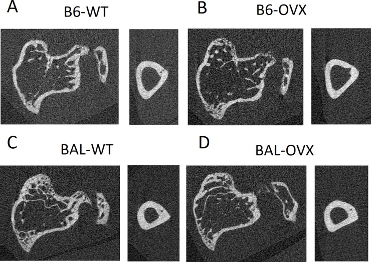
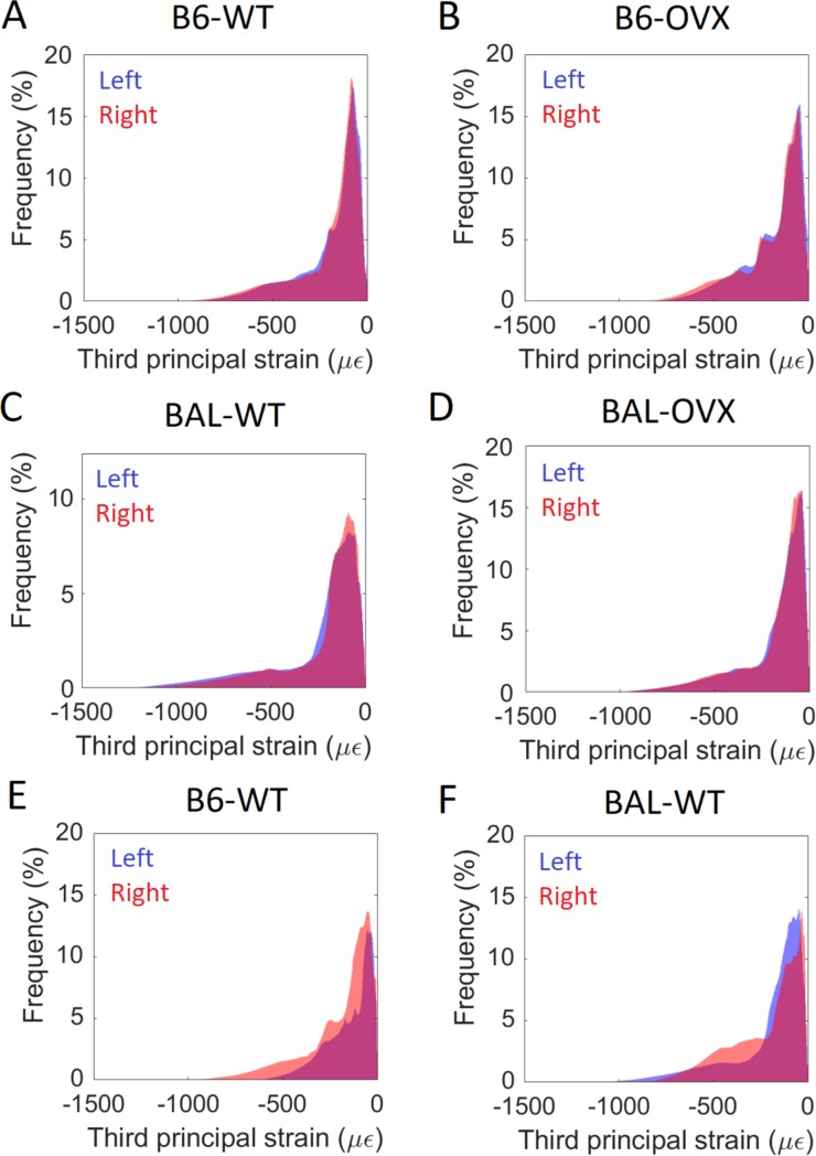
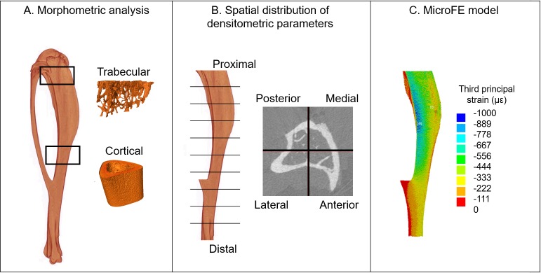
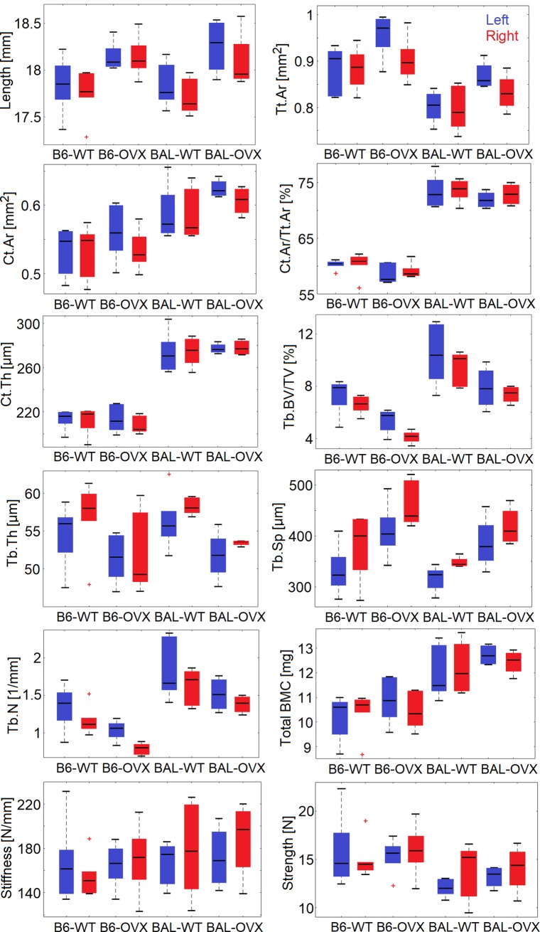
In longitudinal studies, in vivo micro-Computed Tomography (microCT) imaging is used to investigate bone changes over time due to interventions in mice. However, ionising radiation can provoke significant variations in bone morphometric parameters. In a previous study, we evaluated the effect of reducing the integration time on the properties of the mouse tibia measured from microCT images. A scanning procedure (100 ms integration time, 256 mGy nominal radiation dose) was selected as the best compromise between image quality and radiation dose induced on the animal. In this work, the effect of repeated in vivo scans has been evaluated using the selected procedure. The right tibia of twelve female C57BL/6 (six wild type, WT, six ovariectomised, OVX) and twelve BALB/c (six WT, six OVX) mice was scanned every two weeks, starting at week 14 of age. At week 24, mice were sacrificed and both tibiae were scanned. Standard trabecular and cortical morphometric parameters were calculated. The spatial distribution of densitometric parameters (e.g. bone mineral content) was obtained by dividing each tibia in 40 partitions. Stiffness and strength in compression were estimated using homogeneous linear elastic microCT-based micro-Finite Element models. Differences between right (irradiated) and left (non-irradiated control) tibiae were evaluated for each parameter. The irradiated tibiae had higher Tb.Th (+3.3%) and Tb.Sp (+11.6%), and lower Tb.N (-14.2%) compared to non-irradiated tibiae, consistently across both strains and intervention groups. A reduction in Tb.BV/TV (-14.9%) was also observed in the C57BL/6 strain. In the OVX group, a small reduction was also observed in Tt.Ar (-5.0%). In conclusion, repeated microCT scans (at 256 mGy, 5 scans, every two weeks) had limited effects on the mouse tibia, compared to the expected changes induced by bone treatments. Therefore, the selected scanning protocol is acceptable for measuring the effect of bone interventions in vivo.
在纵向研究中,活体微计算机断层扫描(microCT)成像用于研究由于小鼠干预而导致的骨骼随时间的变化。然而,电离辐射会引起骨形态计量参数的显著变化。在之前的一项研究中,我们评估了降低积分时间对从 microCT 图像中测量的小鼠胫骨特性的影响。选择扫描程序(100ms 积分时间,256mGy 名义辐射剂量)作为图像质量和动物辐射剂量之间的最佳折衷方案。在这项工作中,使用选定的程序评估了重复活体扫描的影响。从第 14 周龄开始,每隔两周扫描 12 只雌性 C57BL/6(6 只野生型,WT,6 只卵巢切除,OVX)和 12 只 BALB/c(6 只 WT,6 只 OVX)小鼠的右胫骨。在第 24 周时,处死小鼠并扫描双侧胫骨。计算标准小梁和皮质形态计量参数。通过将每根胫骨分为 40 个分区来获得骨密度参数(如骨矿物质含量)的空间分布。使用均匀线性弹性 microCT 微有限元模型估计压缩时的刚度和强度。对于每个参数,评估右(照射)和左(非照射对照)胫骨之间的差异。与非照射胫骨相比,照射胫骨的 Tb.Th(增加 3.3%)和 Tb.Sp(增加 11.6%)更高,Tb.N(减少 14.2%)更低,两种品系和干预组均如此。在 C57BL/6 品系中还观察到 Tb.BV/TV(减少 14.9%)减少。在 OVX 组中,Tb.Ar(减少 5.0%)也略有减少。总之,与骨处理引起的预期变化相比,重复的 microCT 扫描(在 256mGy,5 次扫描,每两周一次)对小鼠胫骨的影响有限。因此,选定的扫描方案可用于测量体内骨干预的效果。