Kgoebane Kgomotso, Ally Mahmood M T M, Duim-Beytell Martha C, Suleman Farhana E
Department of Radiology, University of Pretoria, South Africa.
Department of Internal Medicine, University of Pretoria, South Africa.
SA J Radiol. 2018 Jul 11;22(1):1316. doi: 10.4102/sajr.v22i1.1316. eCollection 2018.
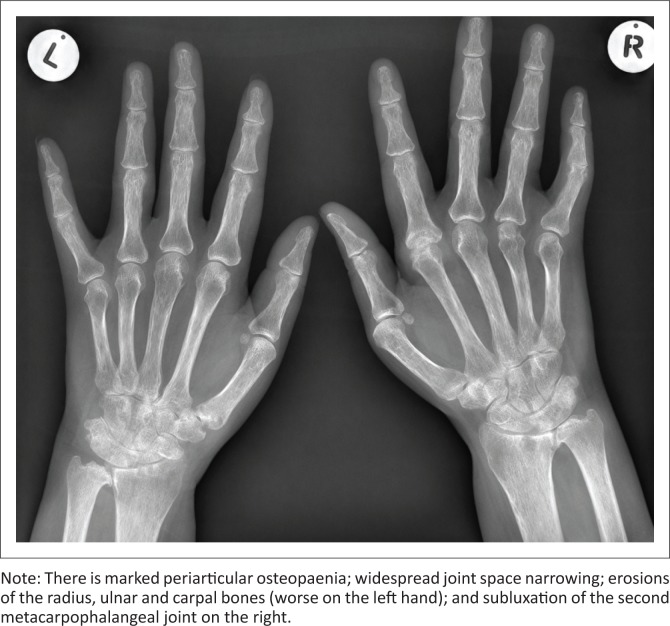
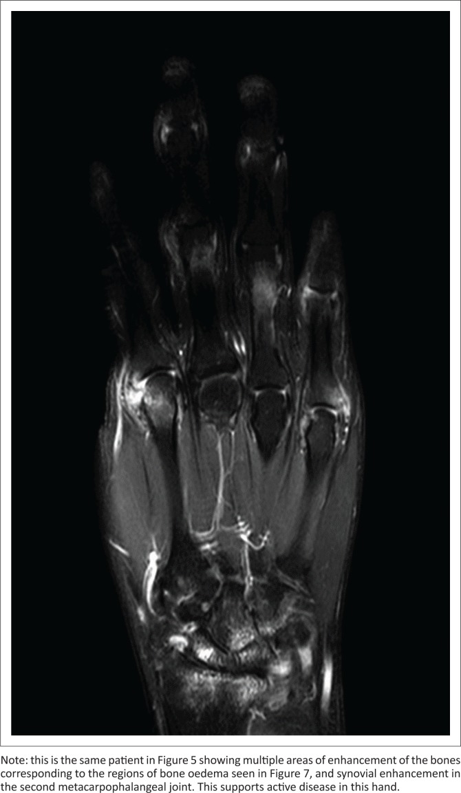
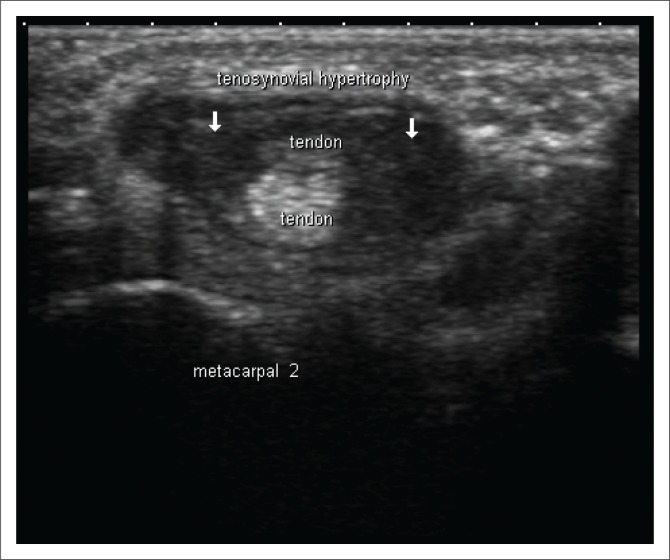
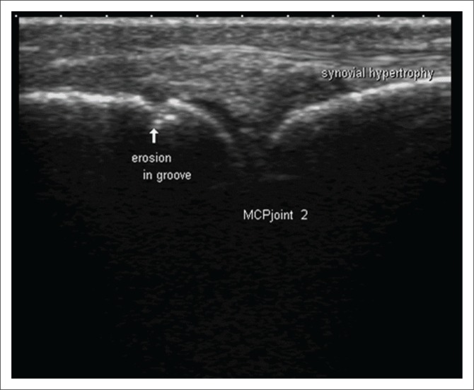
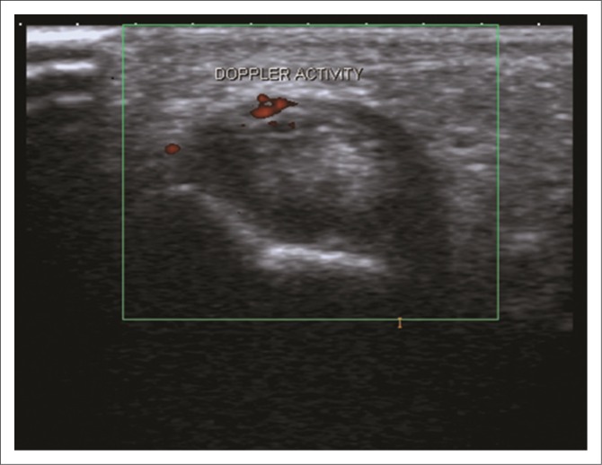
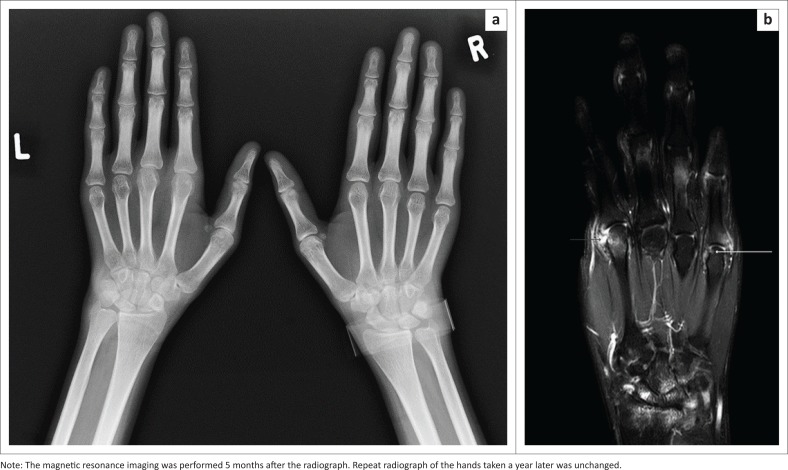
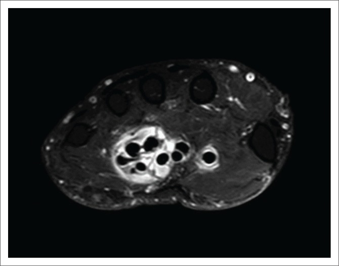
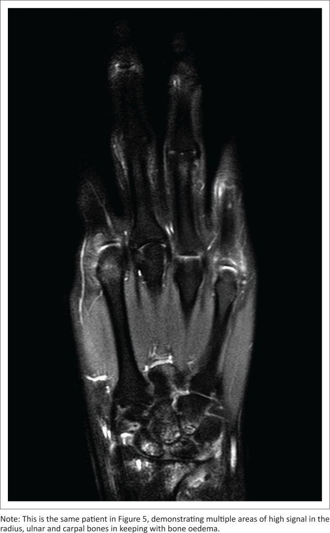
Conventional radiographs of the hands and feet have traditionally been used in the diagnosis, management and monitoring of patients with rheumatoid arthritis (RA). However, they are not sensitive enough to detect changes early in the disease process. Erosions may only be visible up to two years after the onset of disease, and soft tissue involvement may not be detected at all. Early diagnosis can also be made challenging as markers such as erythrocyte sedimentation rate and C-reactive protein may be normal in up to 20% - 25% of cases. The latest classification criteria (American College of Rheumatology/European League Against Rheumatism [ACR/EULAR] Rheumatoid Arthritis Classification criteria 2010), often used to diagnose RA, incorporate the role of ultrasound and magnetic resonance imaging detection of synovitis, enabling earlier diagnosis and correct classification of patients. This article looks at the role of the various imaging modalities used in the diagnosis and management of RA.
传统上,手足的常规X线片一直用于类风湿关节炎(RA)患者的诊断、管理和监测。然而,它们在疾病进程早期检测变化的灵敏度不够。侵蚀可能在疾病发作后长达两年才可见,并且可能根本检测不到软组织受累情况。由于红细胞沉降率和C反应蛋白等标志物在高达20% - 25%的病例中可能正常,早期诊断也可能具有挑战性。最新的分类标准(美国风湿病学会/欧洲抗风湿病联盟[ACR/EULAR]2010年类风湿关节炎分类标准)常用于诊断RA,其中纳入了超声和磁共振成像检测滑膜炎的作用,从而能够对患者进行早期诊断和正确分类。本文探讨了各种成像方式在RA诊断和管理中的作用。