Division of Structural Biology, Wellcome Center for Human Genetics, University of Oxford, OX3 7BN Oxford, United Kingdom.
Laboratory of Virology, National Institute of Allergy and Infectious Diseases, National Institutes of Health, Hamilton, MT 59840.
Proc Natl Acad Sci U S A. 2019 Dec 10;116(50):25057-25067. doi: 10.1073/pnas.1912503116. Epub 2019 Nov 25.
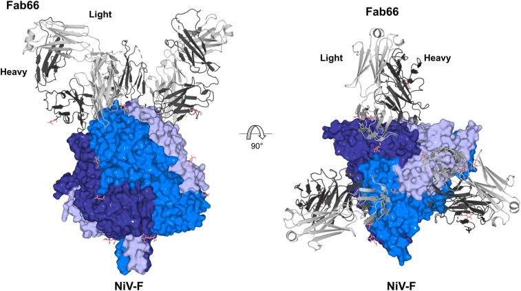
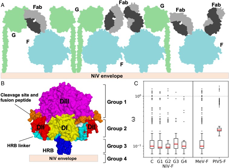
Nipah virus (NiV) is a highly pathogenic paramyxovirus that causes frequent outbreaks of severe neurologic and respiratory disease in humans with high case fatality rates. The 2 glycoproteins displayed on the surface of the virus, NiV-G and NiV-F, mediate host-cell attachment and membrane fusion, respectively, and are targets of the host antibody response. Here, we provide a molecular basis for neutralization of NiV through antibody-mediated targeting of NiV-F. Structural characterization of a neutralizing antibody (nAb) in complex with trimeric prefusion NiV-F reveals an epitope at the membrane-distal domain III (DIII) of the molecule, a region that undergoes substantial refolding during host-cell entry. The epitope of this monoclonal antibody (mAb66) is primarily protein-specific and we observe that glycosylation at the periphery of the interface likely does not inhibit mAb66 binding to NiV-F. Further characterization reveals that a Hendra virus-F-specific nAb (mAb36) and many antibodies in an antihenipavirus-F polyclonal antibody mixture (pAb835) also target this region of the molecule. Integrated with previously reported paramyxovirus F-nAb structures, these data support a model whereby the membrane-distal region of the F protein is targeted by the antibody-mediated immune response across henipaviruses. Notably, our domain-specific sequence analysis reveals no evidence of selective pressure at this region of the molecule, suggestive that functional constraints prevent immune-driven sequence variation. Combined, our data reveal the membrane-distal region of NiV-F as a site of vulnerability on the NiV surface.
寨卡病毒(NiV)是一种高致病性副粘病毒,会导致人类频繁爆发严重的神经和呼吸道疾病,病死率很高。病毒表面展示的 2 种糖蛋白,NiV-G 和 NiV-F,分别介导宿主细胞附着和膜融合,是宿主抗体反应的靶标。在这里,我们通过抗体介导的 NiV-F 靶向作用,为中和 NiV 提供了分子基础。与三聚体预融合 NiV-F 复合物的结构表征揭示了分子膜远端结构域 III(DIII)上的一个中和表位,该区域在宿主细胞进入过程中经历了大量重折叠。该单克隆抗体(mAb66)的表位主要是蛋白特异性的,我们观察到界面周围的糖基化不太可能抑制 mAb66 与 NiV-F 的结合。进一步的表征表明,亨德拉病毒-F 特异性 nAb(mAb36)和抗亨德拉病毒-F 多克隆抗体混合物(pAb835)中的许多抗体也靶向该分子区域。与先前报道的副粘病毒 F-nAb 结构相结合,这些数据支持了一种模型,即抗体介导的免疫反应针对亨尼帕病毒的 F 蛋白的膜远端区域。值得注意的是,我们的特定于结构域的序列分析没有显示该分子区域存在选择压力的证据,这表明功能约束阻止了免疫驱动的序列变异。综合来看,我们的数据揭示了 NiV-F 的膜远端区域是 NiV 表面的一个脆弱部位。