Hofstaetter Jochen G, Misof Barbara M, Jones Dallas C, Zoehrer Ruth, Blouin Stéphane, Schueler Christiane, Paschalis Eleftherios P, Erben Reinhold G, Weinkamer Richard, Klaushofer Klaus, Roschger Paul
1st Medical Department Hanusch Hospital Ludwig Boltzmann Institute of Osteology at the Hanusch Hospital of WGKK and AUVA Trauma Centre Meidling Vienna Austria.
Orthopaedic Hospital Vienna Speising Vienna Austria.
JBMR Plus. 2019 Sep 11;3(11):e10226. doi: 10.1002/jbm4.10226. eCollection 2019 Nov.
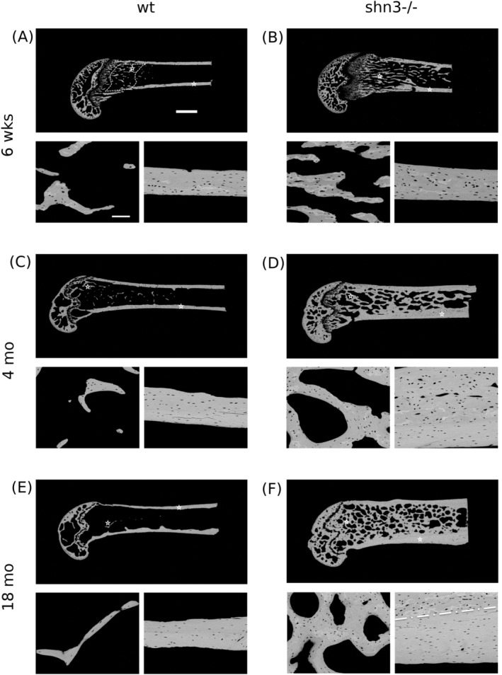
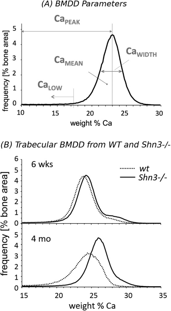
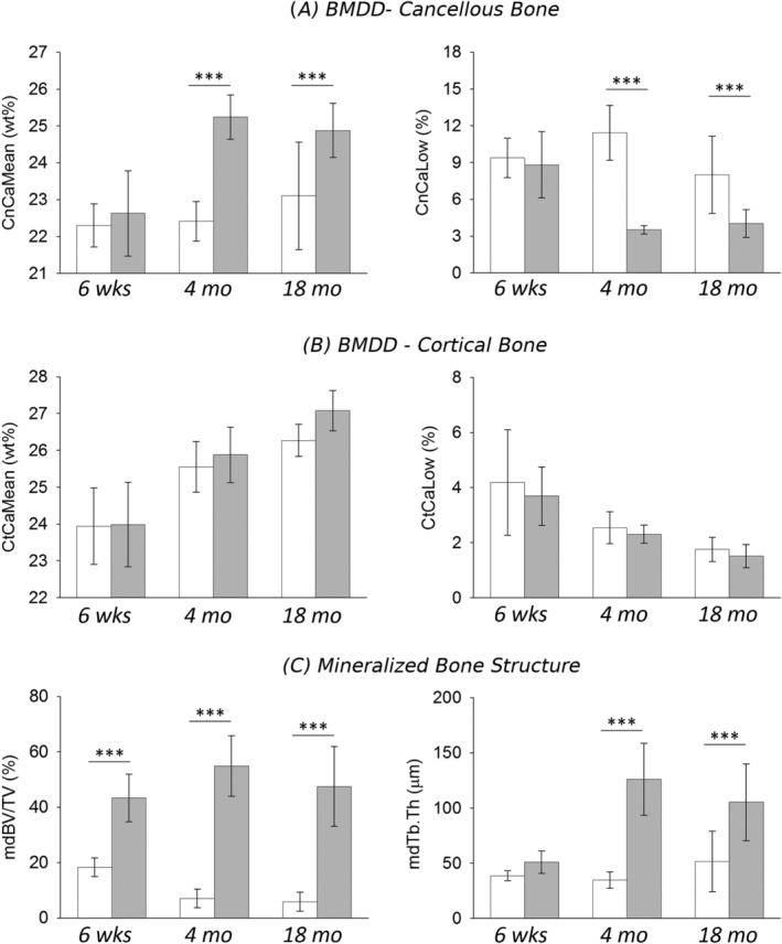
is an essential regulator of postnatal skeletal remodeling. Shn3-deficient mice (Shn3-/-) have high bone mass; however, their bone mechanical and material properties have not been investigated to date. We performed three-point bending of femora, compression tests of L3 vertebrae. We also measured intrinsic material properties, including bone mineralization density distribution (BMDD) and osteocyte lacunae section (OLS) characteristics by quantitative backscatter electron imaging, as well as collagen cross-linking by Fourier transform infrared microspectroscopy of femora from Shn3-/- and WT mice at different ages (6 weeks, 4 months, and 18 months). Moreover, computer modeling was performed for the interpretation of the BMDD outcomes. Femora and L3 vertebrae from Shn3-/- aged 6 weeks revealed increased ultimate force (2.2- and 3.2-fold, < .01, respectively). Mineralized bone volume at the distal femoral metaphysis was about twofold (at 6 weeks) to eightfold (at 4 and 18 months of age) in Shn3-/- ( < .001). Compared with WT, the average degree of trabecular bone mineralization was similar at 6 weeks, but increased at 4 and 18 months of age (+12.6% and +7.7%, < .01, respectively) in Shn3-/-. The analysis of OLS characteristics revealed a higher OLS area for Shn3-/- versus WT at all ages (+16%, +23%, +21%, respectively, < .01). The collagen cross-link ratio was similar between groups. We conclude that femora and vertebrae from Shn3-/- had higher ultimate force in mechanical testing. Computer modeling demonstrated that in cases of highly increased bone volume, the average degree of bone matrix mineralization can be higher than in WT bone, which was actually measured in the older Shn3-/- groups. The area of 2D osteocyte lacunae sections was also increased in Shn3-deficiency, which could only partly be explained by larger remnant areas of primary cortical bone. © 2019 The Authors. published by Wiley Periodicals, Inc. on behalf of American Society for Bone and Mineral Research.
是出生后骨骼重塑的重要调节因子。Shn3基因敲除小鼠(Shn3-/-)骨量较高;然而,其骨力学和材料特性迄今尚未得到研究。我们对股骨进行了三点弯曲试验,对L3椎体进行了压缩试验。我们还通过定量背散射电子成像测量了包括骨矿化密度分布(BMDD)和骨细胞陷窝截面(OLS)特征在内的内在材料特性,以及通过傅里叶变换红外显微光谱法测量了不同年龄(6周、4个月和18个月)的Shn3-/-和野生型(WT)小鼠股骨的胶原交联情况。此外,进行了计算机建模以解释BMDD结果。6周龄的Shn3-/-小鼠的股骨和L3椎体显示出极限力增加(分别增加2.2倍和3.2倍,P<0.01)。Shn3-/-小鼠股骨远端干骺端的矿化骨体积在6周龄时约为两倍(P<0.001),在4个月和18个月龄时约为八倍。与野生型相比,6周龄时小梁骨矿化平均程度相似,但在4个月和18个月龄时Shn3-/-小鼠增加(分别增加12.6%和7.7%,P<0.01)。对OLS特征的分析显示,在所有年龄组中Shn3-/-小鼠的OLS面积均高于野生型(分别增加16%、23%、21%,P<0.01)。各组之间的胶原交联率相似。我们得出结论,在力学测试中,Shn3-/-小鼠的股骨和椎体具有更高的极限力。计算机建模表明,在骨体积高度增加的情况下,骨基质矿化平均程度可能高于野生型骨,这在老年Shn3-/-组中实际得到了测量。Shn3基因缺失时二维骨细胞陷窝截面面积也增加,这只能部分地由初级皮质骨较大的残余面积来解释。©2019作者。由Wiley Periodicals, Inc.代表美国骨与矿物质研究学会出版。