Department of Gastroenterology, Saku Central Hospital Advanced Care Center, 3400-28 Nakagomi, Saku, Nagano, 385-0051, Japan.
Department of Endoscopy, Saku Central Hospital Advanced Care Center, Saku, Nagano, Japan.
Clin J Gastroenterol. 2020 Jun;13(3):365-371. doi: 10.1007/s12328-019-01076-5. Epub 2019 Dec 3.
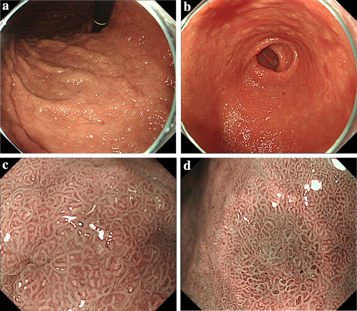
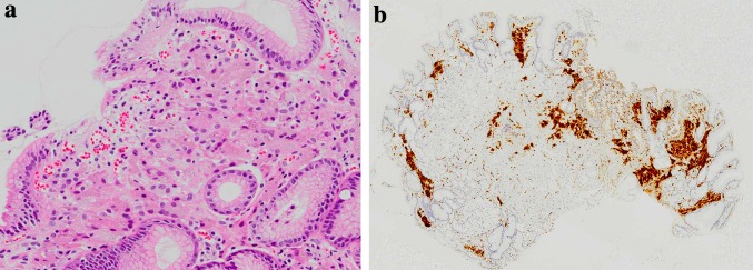
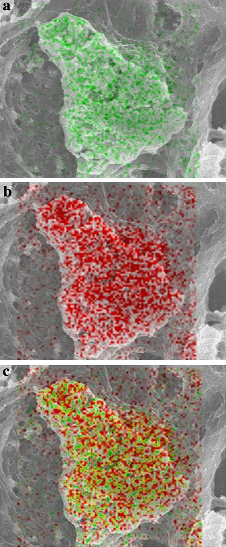
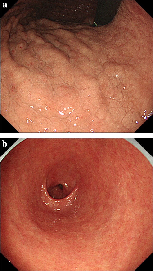
We describe the case of a 70-year-old man with diabetic nephropathy undergoing hemodialysis. Four years following hemodialysis, he started taking lanthanum carbonate 1500 mg/day and lansoprazole 30 mg/day. Nine years following hemodialysis, he underwent screening esophagogastroduodenoscopy, which demonstrated the presence of the whitish cobblestone-like mucosa in the gastric corpus and multiple reddish depressed lesions with annular whitish mucosa in the antrum. With magnified narrow-band imaging endoscopy, a yellowish-white substance was observed in the villous structure, and subepithelial vessels were observed on the yellowish-white substance. Biopsies were taken from the whitish cobblestone-like mucosa of the upper corpus, a reddish depressed part of the antrum. Histologically, aggregates of cells containing amphophilic fine granular material were found in the mucosal interstitium. These cells stained positive for CD68 and were identified as histiocytes. Since he had been taking lanthanum carbonate for 5 years, we considered the possibility of histiocyte-mediated phagocytosis of lanthanum. Digital mapping via scanning electron microscopy with energy-dispersive X-ray spectrometry showed the presence of lanthanum and phosphorus in the interstitium and cytoplasm of histiocytes. The white, rough mucosa in the gastric body appeared 6 months following the commencement of lanthanum administration and still exists 3 years and 5 months after discontinuation of lanthanum.
我们描述了一位 70 岁的男性糖尿病肾病患者的病例,他在接受血液透析 4 年后开始服用碳酸镧 1500mg/天和兰索拉唑 30mg/天。在接受血液透析 9 年后,他进行了筛查性食管胃十二指肠镜检查,发现胃体有白色鹅卵石样黏膜,以及多个红色凹陷性病变,伴有窦部环形白色黏膜。通过放大窄带成像内镜观察,发现绒毛状结构中有淡黄色物质,淡黄色物质上可见上皮下血管。从胃体上部的白色鹅卵石样黏膜和窦部的红色凹陷性部位取活检。组织学上,在黏膜间质中发现含有嗜酸性细颗粒物质的细胞聚集。这些细胞 CD68 染色阳性,被鉴定为组织细胞。由于他已经服用碳酸镧 5 年,我们考虑了组织细胞介导的镧吞噬的可能性。通过扫描电子显微镜和能量色散 X 射线光谱的数字映射显示,组织细胞的间质和细胞质中存在镧和磷。胃体的白色粗糙黏膜在开始服用镧后 6 个月出现,并且在停用镧 3 年零 5 个月后仍然存在。