Mathiasen Anders Bruun, Qayyum Abbas Ali, Jørgensen Erik, Helqvist Steffen, Ekblond Annette, Ng Michael, Bhakoo Kishore, Kastrup Jens
Cardiac Stem Cell Center and Catheterization Laboratory, The Heart Centre, Rigshospitalet, University of Copenhagen, Copenhagen, Denmark.
Translational Imaging Group, Singapore Bioimaging Consortium, Agency for Science, Technology and Research, Singapore, Singapore.
Stem Cells Int. 2019 Nov 14;2019:2754927. doi: 10.1155/2019/2754927. eCollection 2019.
While regenerative stem cell therapy for ischemic heart disease has moved into phase 3 studies, little is still known about retention and migration of cell posttransplantation. In human studies, the ability to track transplanted cells has been limited to labeling with radioisotopes and tracking using nuclear imaging. This method is limited by low resolution and short half-lives of available radioisotopes. Longitudinal tracking using magnetic resonance imaging (MRI) of myocardial injected cells labeled with iron oxide nanoparticles has shown promising results in numerous preclinical studies but has yet to be evaluated in human studies. We aimed to evaluate MRI tracking of mesenchymal stromal cells (MSCs) labeled with ultrasmall paramagnetic iron oxide (USPIO) nanoparticles after intramyocardial transplantation in patients with ischemic heart disease (IHD).
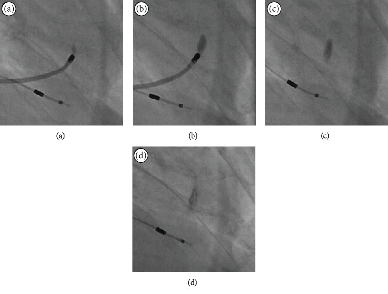
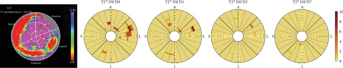
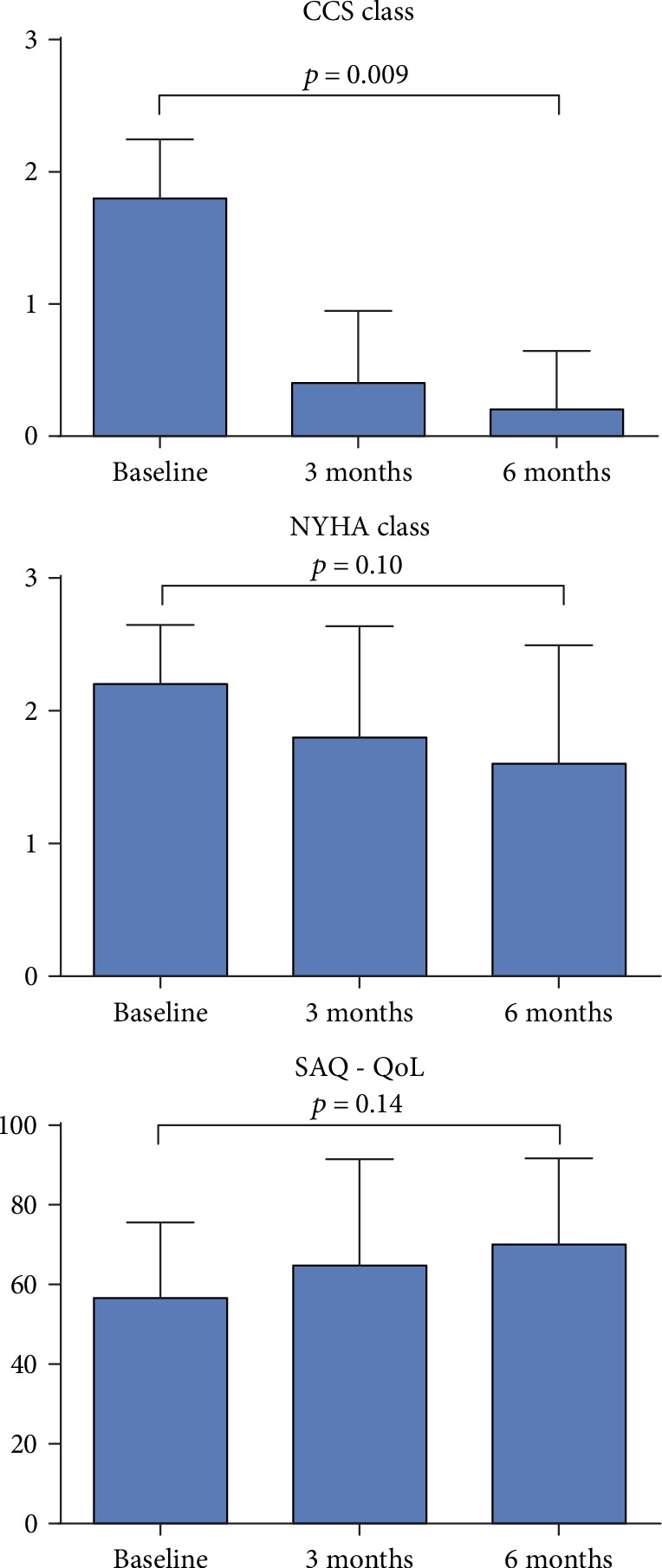
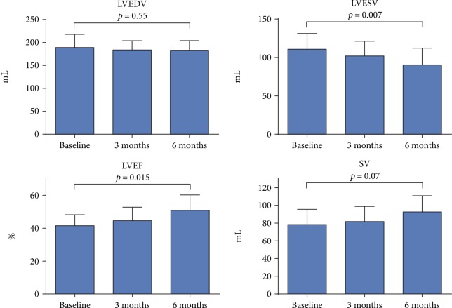
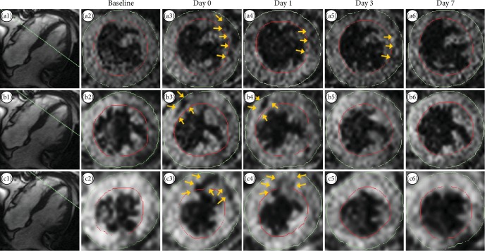
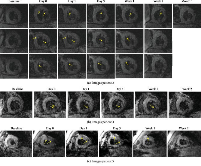
Five no-option patients with chronic symptomatic IHD underwent NOGA-guided intramyocardial transplantation of USPIO-labeled MSCs. Serial MRI scans were performed to track labeled cells both visually and using semiautomated T2∗ relaxation time analysis. For safety, we followed symptoms, quality of life, and myocardial function for 6 months.
USPIO-labeled MSCs were tracked for up to 14 days after transplantation at injection sites both visually and using semiautomated regional T2∗ relaxation time analysis. Labeling of MSCs did not impair long-term safety of treatment.
This was a first-in-man clinical experience aimed at evaluating the utility of MRI tracking of USPIO-labeled bone marrow-derived autologous MSCs after intramyocardial injection in patients with chronic IHD. The treatment was safe, and cells were detectable at injection sites up to 14 days after transplantation. Further studies are needed to clarify if MSCs migrate out of the injection area into other areas of the myocardium or if injected cells are washed out into the peripheral circulation. The trial is registered with ClinicalTrials.gov NCT03651791.
虽然用于缺血性心脏病的再生干细胞疗法已进入3期研究,但对于移植后细胞的留存和迁移仍知之甚少。在人体研究中,追踪移植细胞的能力仅限于用放射性同位素标记并使用核成像进行追踪。这种方法受到可用放射性同位素分辨率低和半衰期短的限制。使用氧化铁纳米颗粒标记心肌注射细胞的磁共振成像(MRI)进行纵向追踪在众多临床前研究中已显示出有前景的结果,但尚未在人体研究中进行评估。我们旨在评估在缺血性心脏病(IHD)患者心肌内移植后,用超小顺磁性氧化铁(USPIO)纳米颗粒标记的间充质基质细胞(MSCs)的MRI追踪情况。
5例有慢性症状的IHD无其他选择的患者接受了在NOGA引导下心肌内移植USPIO标记的MSCs。进行了系列MRI扫描,以通过视觉和半自动T2∗弛豫时间分析追踪标记细胞。为确保安全,我们对症状、生活质量和心肌功能进行了6个月的随访。
在移植后长达14天,通过视觉和半自动区域T2∗弛豫时间分析在注射部位追踪到了USPIO标记的MSCs。MSCs的标记并未损害治疗的长期安全性。
这是首次在人体进行的临床经验,旨在评估在慢性IHD患者心肌内注射后,对USPIO标记的骨髓源性自体MSCs进行MRI追踪的效用。该治疗是安全的,并且在移植后长达14天可在注射部位检测到细胞。需要进一步研究以阐明MSCs是否从注射区域迁移到心肌的其他区域,或者注射的细胞是否被冲洗到外周循环中。该试验已在ClinicalTrials.gov注册,注册号为NCT03651791。