Ugradar Shoaib, Le Alan, Lesgart Michael, Goldberg Robert A, Rootman Daniel, Demer Joseph L
Stein Eye Institute, University of California, Los Angeles, CA, USA.
Bioengineering Interdepartmental Program, University of California, Los Angeles, CA, USA.
Transl Vis Sci Technol. 2019 Dec 5;8(6):25. doi: 10.1167/tvst.8.6.25. eCollection 2019 Nov.
To investigate the feasibility of increasing the stiffness of human tarsal tissue following treatment with riboflavin and ultraviolet A (UVA) to induce cross-linking of collagen fibers.
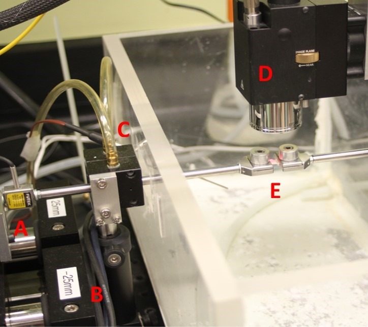
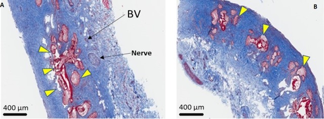
In this case control study, 18 right and left upper eyelids were excised en bloc from 18 fresh-frozen cadavers. One side served as the control while the samples from the opposite side were cross-linked. Four 2 × 6-mm vertical strips of central tarsus were cut from the superior to inferior border of each tarsal plate. Sample tissue was irradiated with UVA at 6 mW/cm for 18 minutes. A microtensile load cell and an optical coherence tomography scanner allowed calculation of stiffness (Young's modulus). Six cross-linked samples and corresponding controls were stained with hematoxylin and eosin (H&E) and Masson trichrome stains. Four controls and four cross-linked samples were also reviewed with a transmission electron microscope.
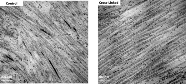
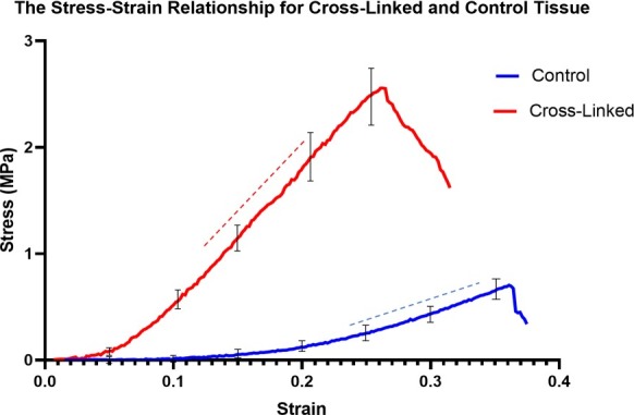
Mean Young's modulus in the linear region for controls was 28 ± 9 MPa and was much higher at 138 ± 8 MPa for cross-linked samples ( < 0.001), yielding a 493% mean stiffness increase. Staining with H&E and Masson did not reveal any histologic changes. Transmission electron microscopy showed a decrease in average diameter of 50 randomly selected collagen fibers from 47.2 ± 1.9 nm prior to cross-linking to 34.2 ± 1.1 nm post cross-linking ( < 0.001). Qualitatively, the collagen fibers appeared more closely packed following cross-linking.
The findings of this study suggest that collagen cross-linking is a viable and effective modality for increasing the stiffness of human tarsal plates.
This work provides proof that collagen cross-linking produces stiffening of the human tarsal plate and may be used in disorders that cause eyelid laxity.
研究用核黄素和紫外线A(UVA)处理以诱导胶原纤维交联后增加人睑板组织硬度的可行性。
在这项病例对照研究中,从18具新鲜冷冻尸体上整块切除18只左右上眼睑。一侧作为对照,另一侧的样本进行交联处理。从每个睑板的上缘至下缘切取四条2×6毫米的中央睑板垂直条带。样本组织用6毫瓦/平方厘米的UVA照射18分钟。使用微拉伸测力传感器和光学相干断层扫描仪计算硬度(杨氏模量)。六个交联样本和相应的对照用苏木精和伊红(H&E)以及马松三色染色法染色。四个对照样本和四个交联样本还用透射电子显微镜进行观察。
对照组线性区域的平均杨氏模量为28±9兆帕,交联样本的平均杨氏模量则高得多,为138±8兆帕(P<0.001),平均硬度增加了493%。H&E和马松染色未显示任何组织学变化。透射电子显微镜显示,随机选择的50根胶原纤维的平均直径从交联前的47.2±1.9纳米降至交联后的34.2±1.1纳米(P<0.001)。定性来看,交联后胶原纤维排列得更紧密。
本研究结果表明,胶原交联是增加人睑板硬度的一种可行且有效的方法。
这项研究证明胶原交联可使人睑板变硬,可用于治疗导致眼睑松弛的疾病。