Department of Biomedical and Pharmaceutical Sciences, University of Montana, 32 Campus Drive, Missoula, MT, 59812, USA.
Center for Biomolecular Structure and Dynamics, University of Montana, 32 Campus Drive, Missoula, MT, 59812, USA.
Nat Commun. 2019 Dec 20;10(1):5825. doi: 10.1038/s41467-019-13768-0.
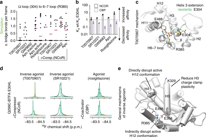
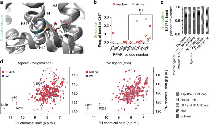
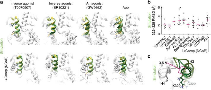
The repressive states of nuclear receptors (i.e., apo or bound to antagonists or inverse agonists) are poorly defined, despite the fact that nuclear receptors are a major drug target. Most ligand bound structures of nuclear receptors, including peroxisome proliferator-activated receptor γ (PPARγ), are similar to the apo structure. Here we use NMR, accelerated molecular dynamics and hydrogen-deuterium exchange mass spectrometry to define the PPARγ structural ensemble. We find that the helix 3 charge clamp positioning varies widely in apo and is stabilized by efficacious ligand binding. We also reveal a previously undescribed mechanism for inverse agonism involving an omega loop to helix switch which induces disruption of a tripartite salt-bridge network. We demonstrate that ligand binding can induce multiple structurally distinct repressive states. One state recruits peptides from two different corepressors, while another recruits just one, providing structural evidence of ligand bias in a nuclear receptor.
核受体的抑制状态(即无配体结合或与拮抗剂或反向激动剂结合)定义不明确,尽管核受体是主要的药物靶点。大多数核受体的配体结合结构,包括过氧化物酶体增殖物激活受体γ(PPARγ),与无配体结合结构相似。在这里,我们使用 NMR、加速分子动力学和氢氘交换质谱来定义 PPARγ 的结构集合。我们发现,无配体结合时,螺旋 3 电荷夹的定位变化很大,而有效配体结合则稳定了它的位置。我们还揭示了一种以前未被描述的反向激动机制,涉及 ω 环到螺旋的转换,这会导致三部分盐桥网络的破坏。我们证明,配体结合可以诱导多种结构上不同的抑制状态。一种状态从两个不同的共抑制因子招募肽,而另一种状态只招募一个,为核受体中的配体偏向提供了结构证据。