• 文献检索
  • 文档翻译
  • 深度研究
  • 学术资讯
  • Suppr Zotero 插件Zotero 插件
  • 邀请有礼
  • 套餐&价格
  • 历史记录
应用&插件
Suppr Zotero 插件Zotero 插件浏览器插件Mac 客户端Windows 客户端微信小程序
定价
高级版会员购买积分包购买API积分包
服务
文献检索文档翻译深度研究API 文档MCP 服务
关于我们
关于 Suppr公司介绍联系我们用户协议隐私条款
关注我们

Suppr 超能文献

核心技术专利:CN118964589B侵权必究
粤ICP备2023148730 号-1Suppr @ 2026

文献检索

告别复杂PubMed语法,用中文像聊天一样搜索,搜遍4000万医学文献。AI智能推荐,让科研检索更轻松。

立即免费搜索

文件翻译

保留排版,准确专业,支持PDF/Word/PPT等文件格式,支持 12+语言互译。

免费翻译文档

深度研究

AI帮你快速写综述,25分钟生成高质量综述,智能提取关键信息,辅助科研写作。

立即免费体验

肠道干细胞衰老受 mTORC1 通过 p38 MAPK-p53 通路驱动。

Gut stem cell aging is driven by mTORC1 via a p38 MAPK-p53 pathway.

机构信息

Bio-X Institutes, Key Laboratory for the Genetics of Developmental and Neuropsychiatric Disorders, Ministry of Education, Shanghai Jiao Tong University, Shanghai, 200240, China.

Department of Biochemistry and Molecular Biology, School of Basic Medical Sciences, Fudan University, Shanghai, 200032, China.

出版信息

Nat Commun. 2020 Jan 2;11(1):37. doi: 10.1038/s41467-019-13911-x.

DOI:10.1038/s41467-019-13911-x
PMID:31896747
原文链接:https://pmc.ncbi.nlm.nih.gov/articles/PMC6940394/
Abstract

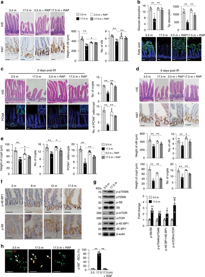
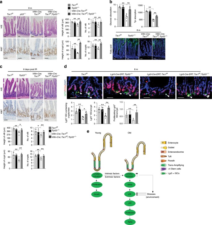
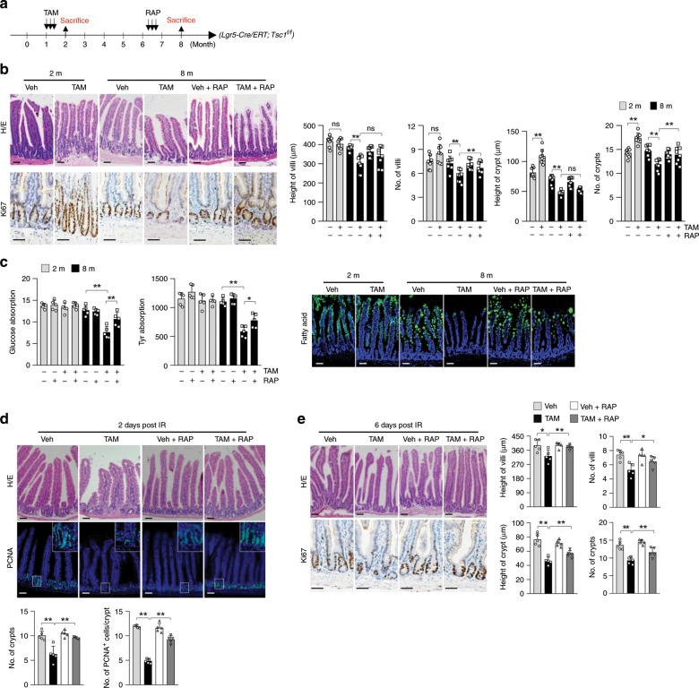
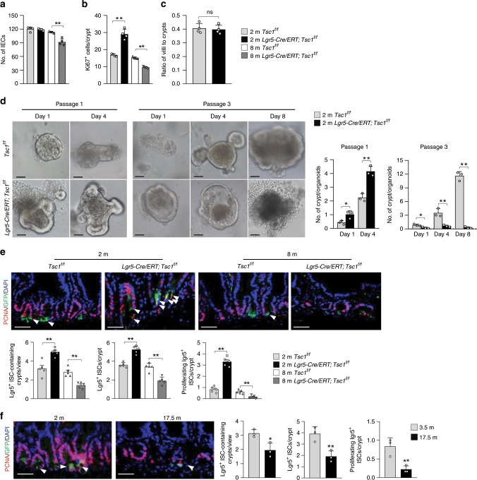
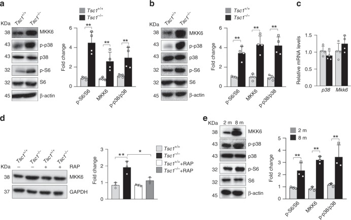
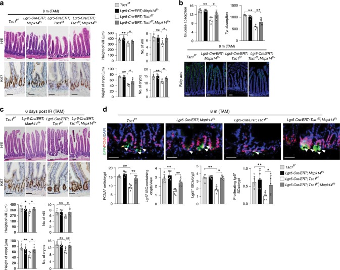
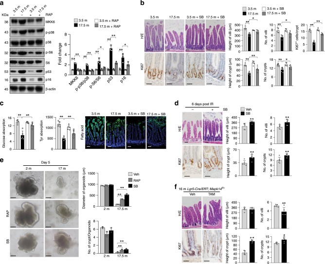
Nutrients are absorbed solely by the intestinal villi. Aging of this organ causes malabsorption and associated illnesses, yet its aging mechanisms remain unclear. Here, we show that aging-caused intestinal villus structural and functional decline is regulated by mTORC1, a sensor of nutrients and growth factors, which is highly activated in intestinal stem and progenitor cells in geriatric mice. These aging phenotypes are recapitulated in intestinal stem cell-specific Tsc1 knockout mice. Mechanistically, mTORC1 activation increases protein synthesis of MKK6 and augments activation of the p38 MAPK-p53 pathway, leading to decreases in the number and activity of intestinal stem cells as well as villus size and density. Targeting p38 MAPK or p53 prevents or rescues ISC and villus aging and nutrient absorption defects. These findings reveal that mTORC1 drives aging by augmenting a prominent stress response pathway in gut stem cells and identify p38 MAPK as an anti-aging target downstream of mTORC1.

摘要

营养物质仅被肠绒毛吸收。该器官的老化会导致吸收不良和相关疾病,但其老化机制尚不清楚。在这里,我们表明,由 mTORC1 调节由衰老引起的肠绒毛结构和功能下降,mTORC1 是营养物质和生长因子的传感器,在老年小鼠的肠干细胞和祖细胞中高度激活。这些衰老表型在肠干细胞特异性 Tsc1 敲除小鼠中得到重现。从机制上讲,mTORC1 的激活增加了 MKK6 的蛋白质合成,并增强了 p38 MAPK-p53 途径的激活,导致肠干细胞数量和活性以及绒毛大小和密度降低。靶向 p38 MAPK 或 p53 可预防或挽救 ISC 和绒毛衰老以及营养吸收缺陷。这些发现表明,mTORC1 通过增强肠道干细胞中突出的应激反应途径来驱动衰老,并确定 p38 MAPK 是 mTORC1 下游的抗衰老靶点。

https://cdn.ncbi.nlm.nih.gov/pmc/blobs/6020/6940394/def63d323463/41467_2019_13911_Fig7_HTML.jpg
https://cdn.ncbi.nlm.nih.gov/pmc/blobs/6020/6940394/8fcb69ec2109/41467_2019_13911_Fig1_HTML.jpg
https://cdn.ncbi.nlm.nih.gov/pmc/blobs/6020/6940394/baacbd5a6f2d/41467_2019_13911_Fig2_HTML.jpg
https://cdn.ncbi.nlm.nih.gov/pmc/blobs/6020/6940394/a25d74ebec15/41467_2019_13911_Fig3_HTML.jpg
https://cdn.ncbi.nlm.nih.gov/pmc/blobs/6020/6940394/c013935b7582/41467_2019_13911_Fig4_HTML.jpg
https://cdn.ncbi.nlm.nih.gov/pmc/blobs/6020/6940394/48abd8537047/41467_2019_13911_Fig5_HTML.jpg
https://cdn.ncbi.nlm.nih.gov/pmc/blobs/6020/6940394/bb0751483353/41467_2019_13911_Fig6_HTML.jpg
https://cdn.ncbi.nlm.nih.gov/pmc/blobs/6020/6940394/def63d323463/41467_2019_13911_Fig7_HTML.jpg

相似文献

1
Gut stem cell aging is driven by mTORC1 via a p38 MAPK-p53 pathway.肠道干细胞衰老受 mTORC1 通过 p38 MAPK-p53 通路驱动。
Nat Commun. 2020 Jan 2;11(1):37. doi: 10.1038/s41467-019-13911-x.
2
mTORC1 promotes mineralization via p53 pathway.mTORC1 通过 p53 通路促进矿化。
FASEB J. 2021 Feb;35(2):e21325. doi: 10.1096/fj.202002016R.
3
l-Glutamate drives porcine intestinal epithelial renewal by increasing stem cell activity via upregulation of the EGFR-ERK-mTORC1 pathway.l-谷氨酸通过上调 EGFR-ERK-mTORC1 通路增加干细胞活性来驱动猪肠道上皮细胞更新。
Food Funct. 2020 Mar 26;11(3):2714-2724. doi: 10.1039/c9fo03065d.
4
PP2Cdelta (Ppm1d, WIP1), an endogenous inhibitor of p38 MAPK, is regulated along with Trp53 and Cdkn2a following p38 MAPK inhibition during mouse preimplantation development.PP2Cδ(Ppm1d,WIP1)是p38丝裂原活化蛋白激酶(p38 MAPK)的一种内源性抑制剂,在小鼠植入前发育过程中,p38 MAPK受到抑制后,它与Trp53和Cdkn2a一起受到调控。
Mol Reprod Dev. 2007 Jul;74(7):821-34. doi: 10.1002/mrd.20688.
5
Metformin potentiates the effect of arsenic trioxide suppressing intrahepatic cholangiocarcinoma: roles of p38 MAPK, ERK3, and mTORC1.二甲双胍增强三氧化二砷抑制肝内胆管癌的作用:p38丝裂原活化蛋白激酶、ERK3和雷帕霉素靶蛋白复合物1的作用
J Hematol Oncol. 2017 Feb 28;10(1):59. doi: 10.1186/s13045-017-0424-0.
6
Osteocyte TSC1 promotes sclerostin secretion to restrain osteogenesis in mice.破骨细胞 TSC1 促进骨钙素分泌,从而抑制小鼠成骨。
Open Biol. 2019 May 31;9(5):180262. doi: 10.1098/rsob.180262.
7
mTORC1 signaling activation increases intestinal stem cell activity and promotes epithelial cell proliferation.mTORC1 信号激活可增加肠道干细胞活性,促进上皮细胞增殖。
J Cell Physiol. 2019 Aug;234(10):19028-19038. doi: 10.1002/jcp.28542. Epub 2019 Apr 1.
8
Tuberous sclerosis complex-mediated mTORC1 overactivation promotes age-related hearing loss.结节性硬化症复合物介导的 mTORC1 过度激活促进年龄相关性听力损失。
J Clin Invest. 2018 Nov 1;128(11):4938-4955. doi: 10.1172/JCI98058. Epub 2018 Sep 24.
9
TSC1 deletion in fibroblasts alleviates lipopolysaccharide-induced acute kidney injury.成纤维细胞中 TSC1 缺失可减轻脂多糖诱导的急性肾损伤。
Clin Sci (Lond). 2018 Oct 2;132(19):2087-2101. doi: 10.1042/CS20180348. Print 2018 Oct 15.
10
Clostridium difficile toxin A-induced colonocyte apoptosis involves p53-dependent p21(WAF1/CIP1) induction via p38 mitogen-activated protein kinase.艰难梭菌毒素A诱导的结肠上皮细胞凋亡涉及通过p38丝裂原活化蛋白激酶的p53依赖性p21(WAF1/CIP1)诱导。
Gastroenterology. 2005 Dec;129(6):1875-88. doi: 10.1053/j.gastro.2005.09.011.

引用本文的文献

1
Roles of intestinal stem cells in inflammatory bowel disease pathogenesis.肠道干细胞在炎症性肠病发病机制中的作用。
World J Stem Cells. 2025 Aug 26;17(8):107639. doi: 10.4252/wjsc.v17.i8.107639.
2
Crosstalk between dysregulated amino acid sensing and glucose and lipid metabolism in colorectal cancer.结直肠癌中失调的氨基酸感知与葡萄糖和脂质代谢之间的相互作用。
Front Oncol. 2025 Aug 29;15:1665056. doi: 10.3389/fonc.2025.1665056. eCollection 2025.
3
Comprehensive analysis of keloid super-enhancer networks reveals FOXP1-mediated anti-senescence mechanisms in fibrosis.

本文引用的文献

1
Fasting Activates Fatty Acid Oxidation to Enhance Intestinal Stem Cell Function during Homeostasis and Aging.禁食激活脂肪酸氧化以增强稳态和衰老过程中的肠道干细胞功能。
Cell Stem Cell. 2018 May 3;22(5):769-778.e4. doi: 10.1016/j.stem.2018.04.001.
2
Calorie Restriction Governs Intestinal Epithelial Regeneration through Cell-Autonomous Regulation of mTORC1 in Reserve Stem Cells.热量限制通过储备干细胞中 mTORC1 的细胞自主调节来控制肠道上皮细胞再生。
Stem Cell Reports. 2018 Mar 13;10(3):703-711. doi: 10.1016/j.stemcr.2018.01.026. Epub 2018 Mar 1.
3
mTORC1 Activation during Repeated Regeneration Impairs Somatic Stem Cell Maintenance.
瘢痕疙瘩超级增强子网络的综合分析揭示了FOXP1介导的纤维化抗衰老机制。
Cell Mol Biol Lett. 2025 Jul 23;30(1):88. doi: 10.1186/s11658-025-00763-1.
4
Trends in intestinal aging: From underlying mechanisms to therapeutic strategies.肠道衰老的趋势:从潜在机制到治疗策略。
Acta Pharm Sin B. 2025 Jul;15(7):3372-3403. doi: 10.1016/j.apsb.2025.05.011. Epub 2025 May 22.
5
Lipoic acid functions in Paneth cells to prevent human intestinal stem cell aging.硫辛酸在潘氏细胞中发挥作用,以防止人类肠道干细胞衰老。
Nat Commun. 2025 Jul 1;16(1):6016. doi: 10.1038/s41467-025-61070-z.
6
Unraveling Botulinum Neurotoxin A Light-Chain-Induced Signaling Pathways: A Phosphoproteomic Analysis in a Controlled Cellular Model.解析肉毒杆菌神经毒素A轻链诱导的信号通路:在可控细胞模型中的磷酸化蛋白质组学分析
Int J Mol Sci. 2025 May 28;26(11):5168. doi: 10.3390/ijms26115168.
7
Cell cycle regulation by the ribotoxic stress response.核糖体毒性应激反应对细胞周期的调控。
Trends Cell Biol. 2025 Jul;35(7):592-603. doi: 10.1016/j.tcb.2025.04.005. Epub 2025 May 16.
8
Dapagliflozin ameliorates intestinal stem cell aging by regulating the MAPK signaling pathway in .达格列净通过调节……中的丝裂原活化蛋白激酶信号通路改善肠道干细胞衰老。
Front Cell Dev Biol. 2025 Apr 23;13:1576258. doi: 10.3389/fcell.2025.1576258. eCollection 2025.
9
Cow Placenta Peptides Ameliorate D-Galactose-Induced Intestinal Barrier Damage by Regulating TLR/NF-κB Pathway.牛胎盘肽通过调节TLR/NF-κB信号通路改善D-半乳糖诱导的肠道屏障损伤
Vet Sci. 2025 Mar 3;12(3):229. doi: 10.3390/vetsci12030229.
10
Alternate Day Fasting Enhances Intestinal Epithelial Function During Aging by Regulating Mitochondrial Metabolism.隔日禁食通过调节线粒体代谢增强衰老过程中的肠道上皮功能。
Aging Cell. 2025 Jul;24(7):e70052. doi: 10.1111/acel.70052. Epub 2025 Apr 1.
反复再生过程中 mTORC1 的激活会损害体干细胞的维持。
Cell Stem Cell. 2017 Dec 7;21(6):806-818.e5. doi: 10.1016/j.stem.2017.11.008.
4
An Amazing Turn of Events.世事难料。
Cell. 2017 Sep 21;171(1):18-22. doi: 10.1016/j.cell.2017.08.021. Epub 2017 Sep 6.
5
Hierarchy and Plasticity in the Intestinal Stem Cell Compartment.肠道干细胞龛中的层次结构和可塑性。
Trends Cell Biol. 2017 Oct;27(10):753-764. doi: 10.1016/j.tcb.2017.06.006. Epub 2017 Jul 18.
6
In Vivo Imaging Reveals Existence of Crypt Fission and Fusion in Adult Mouse Intestine.体内成像揭示成年小鼠肠道中隐窝裂变和融合的存在。
Gastroenterology. 2017 Sep;153(3):674-677.e3. doi: 10.1053/j.gastro.2017.05.019. Epub 2017 May 26.
7
mTOR Signaling in Growth, Metabolism, and Disease.生长、代谢及疾病中的mTOR信号传导
Cell. 2017 Apr 6;169(2):361-371. doi: 10.1016/j.cell.2017.03.035.
8
Targeted Apoptosis of Senescent Cells Restores Tissue Homeostasis in Response to Chemotoxicity and Aging.衰老细胞的靶向凋亡可恢复组织稳态以应对化学毒性和衰老。
Cell. 2017 Mar 23;169(1):132-147.e16. doi: 10.1016/j.cell.2017.02.031.
9
Canonical Wnt Signaling Ameliorates Aging of Intestinal Stem Cells.经典Wnt信号通路可改善肠道干细胞衰老。
Cell Rep. 2017 Mar 14;18(11):2608-2621. doi: 10.1016/j.celrep.2017.02.056.
10
Interplay between metabolic identities in the intestinal crypt supports stem cell function.肠道隐窝中的代谢身份相互作用支持干细胞功能。
Nature. 2017 Mar 16;543(7645):424-427. doi: 10.1038/nature21673. Epub 2017 Mar 8.