Centre for Osteoarthritis Pathogenesis Versus Arthritis, Kennedy Institute of Rheumatology, Roosevelt Drive, Nuffield Department of Orthopaedics, Rheumatology and Musculoskeletal Sciences, University of Oxford, UK.
Centre for Osteoarthritis Pathogenesis Versus Arthritis, Kennedy Institute of Rheumatology, Nuffield Department of Orthopaedics, Rheumatology and Musculoskeletal Sciences, University of Oxford, UK.
Osteoarthritis Cartilage. 2020 Mar;28(3):324-333. doi: 10.1016/j.joca.2019.12.005. Epub 2020 Jan 2.
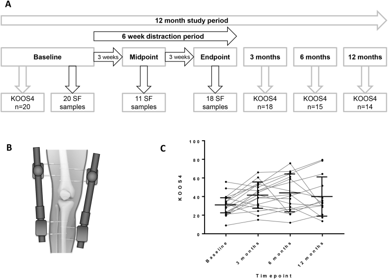
Surgical knee joint distraction (KJD) leads to clinical improvement in knee osteoarthritis (OA) and also apparent cartilage regeneration by magnetic resonance imaging. We investigated if alteration of the joint's mechanical environment during the 6 week period of KJD was associated with a molecular response in synovial fluid, and if any change was associated with clinical response.
20 individuals undergoing KJD for symptomatic radiographic knee OA had SF sampled at baseline, midpoint and endpoint of distraction (6 weeks). SF supernatants were measured by immunoassay for 10 predefined mechanosensitive molecules identified in our previous pre-clinical studies. The composite Knee injury and OA Outcome Score-4 (KOOS) was collected at baseline, 3, 6 and 12 months.
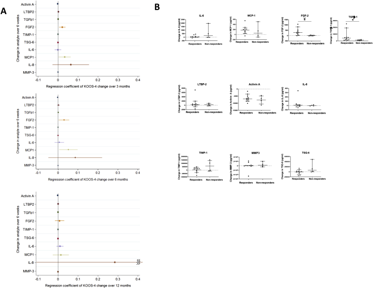
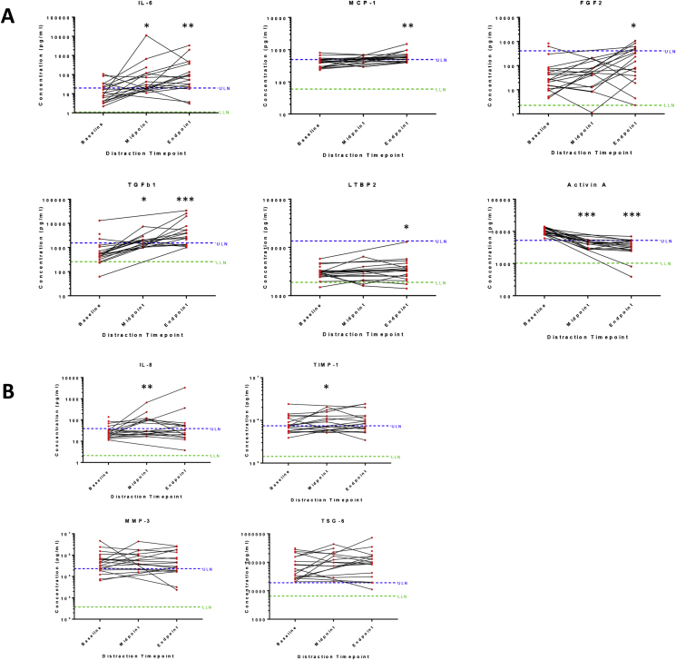
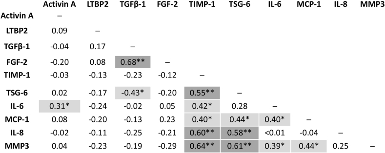
13/20 (65%) were male with mean age 54°±°5yrs. All had Kellgren-Lawrence grade ≥2 knee OA. 6/10 analytes showed statistically significant change in SF over the 6 weeks distraction (activin A; TGFβ-1; MCP-1; IL-6; FGF-2; LTBP2), P < 0.05. Of these, all but activin A increased. Those achieving the minimum clinically important difference of 10 points for KOOS over 6 months showed greater increases in FGF-2 and TGFβ-1 than non-responders. An increase in IL-8 during the 6 weeks of KJD was associated with significantly greater improvement in KOOS over 12 months.
Detectable, significant molecular changes are observed in SF following KJD, that are remarkably consistent between individuals. Preliminary findings appear to suggest that increases in some molecules are associated with clinically meaningful responses. Joint distraction may provide a potential opportunity in the future to define regenerative biomarker(s) and identify pathways that drive intrinsic cartilage repair.
膝关节手术牵张(KJD)可改善膝关节骨关节炎(OA)的临床症状,并通过磁共振成像明显促进软骨再生。我们研究了在 KJD 的 6 周期间关节力学环境的改变是否与滑液中的分子反应相关,以及任何改变是否与临床反应相关。
20 名接受 KJD 治疗的症状性放射学膝关节 OA 患者在基线、牵张中点和终点(6 周)采集滑液样本。通过免疫测定法测量滑液上清液中的 10 种先前在临床前研究中确定的机械敏感分子。在基线、3、6 和 12 个月时收集综合膝关节损伤和骨关节炎结局评分-4(KOOS)。
20 名患者中 13 名(65%)为男性,平均年龄 54°±°5 岁。所有患者均有 Kellgren-Lawrence 分级≥2 级的膝关节 OA。在 6 周的 KJD 期间,有 6/10 种分析物在 SF 中显示出统计学上的显著变化(激活素 A;TGFβ-1;MCP-1;IL-6;FGF-2;LTBP2),P<0.05。其中,除激活素 A 外,所有物质均增加。在 6 个月时 KOOS 达到最小临床重要差异 10 分的患者,其 FGF-2 和 TGFβ-1 的增加幅度大于无反应者。在 6 周的 KJD 期间,IL-8 的增加与 KOOS 在 12 个月内的显著改善相关。
在 KJD 后,SF 中观察到可检测的、显著的分子变化,个体之间非常一致。初步发现表明,某些分子的增加与有临床意义的反应相关。关节牵张术可能为未来定义再生生物标志物和确定驱动内在软骨修复的途径提供潜在机会。