Zhuang You-Yuan, Xiang Lue, Wen Xin-Ran, Shen Ren-Juan, Zhao Ning, Zheng Si-Si, Han Ru-Yi, Qu Jia, Lu Fan, Jin Zi-Bing
Division of Ophthalmic Genetics, The Eye Hospital, Wenzhou Medical University, Wenzhou, China.
State Key Laboratory of Ophthalmology, Optometry and Visual Science, National Clinical Research Center for Ophthalmology, National Center for International Research in Regenerative Medicine and Neurogenetics, Wenzhou, China.
Front Cell Dev Biol. 2019 Dec 12;7:333. doi: 10.3389/fcell.2019.00333. eCollection 2019.
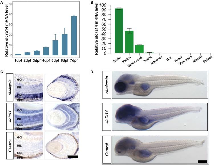
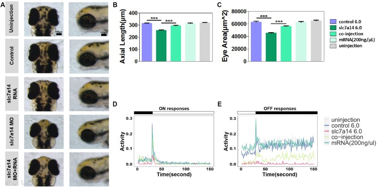
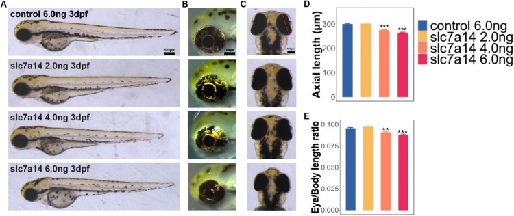
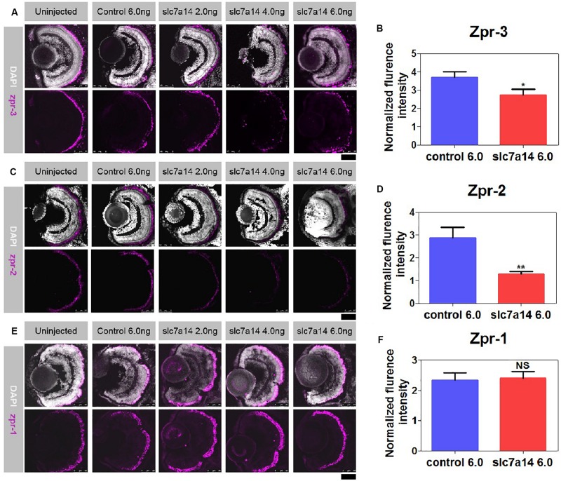
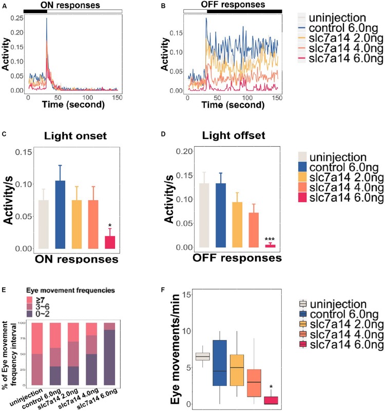
Previous study has identified as a new causative gene of retinitis pigmentosa (RP). However, the role of has not been fully characterized. The goal of this study was to investigate the biological features of in zebrafish. To determine the expression of in developing zebrafish, we performed hybridization (ISH) and quantitative real-time PCR. Morpholino knockdown and overexpression experiments were performed to study the role of in zebrafish retinas. Immunostaining was carried out to observe structural changes. Visual motor responses (VMR) and optokinetic responses (OKR) were analyzed to assess visual behaviors. Terminal deoxynucleotidyl transferase (dUTP) nick-end labeling (TUNEL) staining was performed to survey apoptotic retinal cells. We found that was highly expressed in neuronal tissues, including the brain, spinal cord and retina, and that the expression levels increased during early embryogenesis. Consistently, ISH showed a similar expression pattern. Knockdown of led to dose-dependent microphthalmia that was reversed by overexpression. The immunostaining results revealed that the rod-specific protein zpr-3 and the retinal pigment epithelium-specific protein zpr-2 (decreased to 44.48%) were significantly suppressed in the -silenced morphants. Notably, visual behaviors (the VMR and the OKR) were severely impaired in the -deficient morphant, especially the VMR OFF response. In addition, apoptotic cells were observed in the retina at 3 days post fertilization (dpf) and 5 dpf by TUNEL assay. Our results demonstrated that is essential for visually mediated behaviors in zebrafish. Temporary silencing of in larvae led to severe visual impairments, consistent with the manifestations observed in RP patients. Our findings provide further insights into the genetic mechanisms of RP predisposition caused by mutations.
先前的研究已确定某基因是视网膜色素变性(RP)的一个新致病基因。然而,该基因的作用尚未完全明确。本研究的目的是探究该基因在斑马鱼中的生物学特性。为了确定该基因在发育中的斑马鱼中的表达情况,我们进行了原位杂交(ISH)和定量实时PCR。进行了吗啉代敲低和过表达实验,以研究该基因在斑马鱼视网膜中的作用。进行免疫染色以观察结构变化。分析视觉运动反应(VMR)和视动反应(OKR)以评估视觉行为。进行末端脱氧核苷酸转移酶(dUTP)缺口末端标记(TUNEL)染色以检测视网膜凋亡细胞。我们发现该基因在包括脑、脊髓和视网膜在内的神经组织中高表达,且在胚胎早期发育过程中表达水平升高。一致地,ISH显示出相似的表达模式。该基因的敲低导致剂量依赖性小眼症,而过表达可逆转此现象。免疫染色结果显示,在该基因沉默的 morphants 中,视杆细胞特异性蛋白 zpr - 3 和视网膜色素上皮特异性蛋白 zpr - 2(降至44.48%)显著受到抑制。值得注意的是,在该基因缺陷的 morphant 中,视觉行为(VMR 和 OKR)严重受损,尤其是 VMR OFF 反应。此外,通过 TUNEL 检测在受精后3天(dpf)和5 dpf 的视网膜中观察到凋亡细胞。我们的结果表明,该基因对斑马鱼视觉介导的行为至关重要。幼虫期该基因的暂时沉默导致严重的视觉障碍,这与在RP患者中观察到的表现一致。我们的发现为该基因突变导致RP易感性的遗传机制提供了进一步的见解。