• 文献检索
  • 文档翻译
  • 深度研究
  • 学术资讯
  • Suppr Zotero 插件Zotero 插件
  • 邀请有礼
  • 套餐&价格
  • 历史记录
应用&插件
Suppr Zotero 插件Zotero 插件浏览器插件Mac 客户端Windows 客户端微信小程序
定价
高级版会员购买积分包购买API积分包
服务
文献检索文档翻译深度研究API 文档MCP 服务
关于我们
关于 Suppr公司介绍联系我们用户协议隐私条款
关注我们

Suppr 超能文献

核心技术专利:CN118964589B侵权必究
粤ICP备2023148730 号-1Suppr @ 2026

文献检索

告别复杂PubMed语法,用中文像聊天一样搜索,搜遍4000万医学文献。AI智能推荐,让科研检索更轻松。

立即免费搜索

文件翻译

保留排版,准确专业,支持PDF/Word/PPT等文件格式,支持 12+语言互译。

免费翻译文档

深度研究

AI帮你快速写综述,25分钟生成高质量综述,智能提取关键信息,辅助科研写作。

立即免费体验

共存的脑血管粥样硬化可预测随后的血管事件:一项多对比度心血管磁共振成像研究。

Co-existing cerebrovascular atherosclerosis predicts subsequent vascular event: a multi-contrast cardiovascular magnetic resonance imaging study.

机构信息

Department of Radiology, The Affiliated BenQ Hospital of Nanjing Medical University, Nanjing, China.

Center for Biomedical Imaging Research, Department of Biomedical Engineering, Tsinghua University School of Medicine, Haidian District, Beijing, 100084, China.

出版信息

J Cardiovasc Magn Reson. 2020 Jan 13;22(1):4. doi: 10.1186/s12968-019-0596-6.

DOI:10.1186/s12968-019-0596-6
PMID:31928532
原文链接:https://pmc.ncbi.nlm.nih.gov/articles/PMC6956475/
Abstract

BACKGROUND

It is still unknown that whether co-existing intracranial stenosis and extracranial carotid vulnerable plaques have higher predictive value for subsequent vascular events. This study aimed to determine the relationship between co-existing extracranial carotid vulnerable plaques and intracranial stenosis and subsequent vascular events utilizing cardiovascular magnetic resonance (CMR) vessel wall imaging.

METHODS

Patients who had recent cerebrovascular symptoms in anterior circulation (< 2 weeks) were consecutively enrolled and underwent multi-contrast CMR vessel wall imaging for extracranial carotid arteries and 3D time-of flight CMR angiography for intracranial arteries at baseline. After baseline examination, all patients were followed-up for at least 1 year to determined recurrence of vascular events. The co-existing cerebrovascular atherosclerosis was defined as presence of both intracranial artery stenosis and at least one the following measures of extracranial artery atherosclerosis: plaque, calcification, lipid-rich necrotic core (LRNC), or intraplaque hemorrhage. Univariate and multivariate Cox regressions were used to calculate the hazard ratio (HR) and corresponding 95% confidence interval (CI) of co-existing plaques in predicting subsequent vascular events.

RESULTS

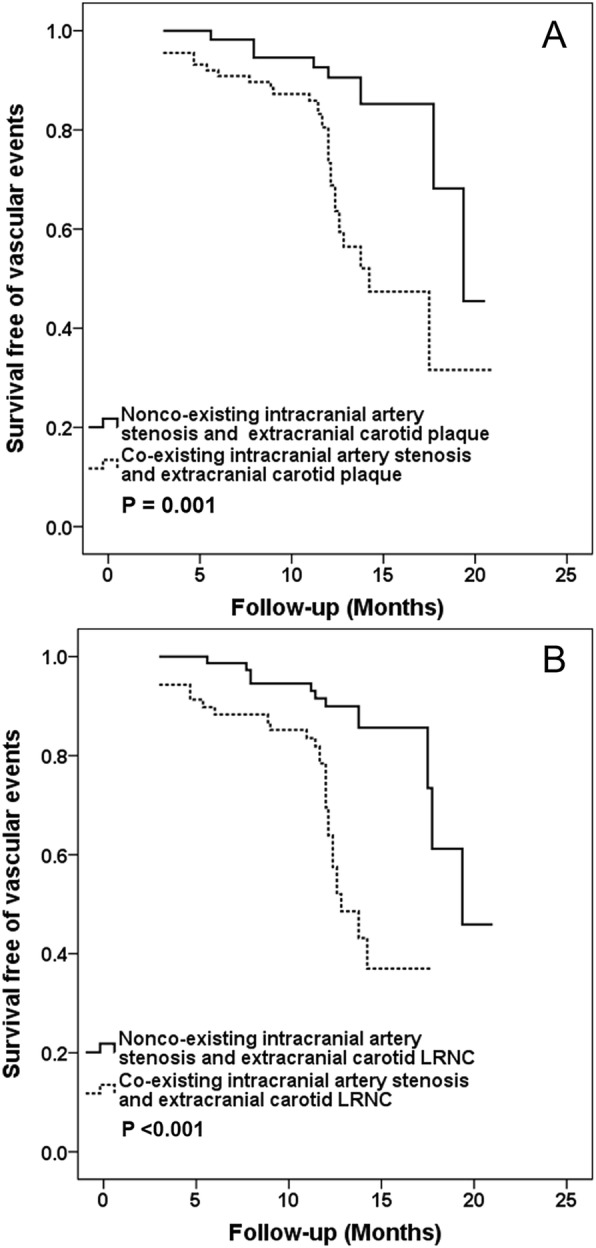
In total, 150 patients (mean age: 61.8 ± 11.9 years; 109 males) were recruited. During the median follow-up time of 12.1 months, 41 (27.3%) patients experienced vascular events. Co-existing intracranial artery stenosis and extracranial carotid plaque (HR, 3.57; 95% CI, 1.63-7.82; P = 0.001) and co-existing intracranial artery stenosis and extracranial carotid LRNC (HR, 4.47; 95% CI, 2.15-9.27; P < 0.001) were significantly associated with subsequent vascular events, respectively. After adjusted for confounding factors and carotid stenosis, these associations remained statistically significant (HR, 5.12; 95% CI, 1.36-19.24; P = 0.016 and HR, 8.12; 95% CI, 2.41-27.31; P = 0.001, respectively).

CONCLUSIONS

The co-existing cerebrovascular atherosclerotic diseases, particularly co-existing carotid lipid-rich necrotic core and intracranial stenosis, are independent predictors for subsequent vascular events.

摘要

背景

目前尚不清楚同时存在颅内狭窄和颅外颈动脉易损斑块是否对后续血管事件具有更高的预测价值。本研究旨在利用心血管磁共振(CMR)血管壁成像来确定颅外颈动脉易损斑块与颅内狭窄并存与后续血管事件之间的关系。

方法

连续纳入近期在前循环(<2 周)出现脑血管症状的患者,并在基线时进行多对比 CMR 血管壁成像以评估颅外颈动脉,以及 3D 时间飞跃 CMR 血管造影以评估颅内动脉。在基线检查后,所有患者均至少随访 1 年以确定血管事件的复发情况。共存性脑血管粥样硬化定义为同时存在颅内动脉狭窄和以下至少一项颅外动脉粥样硬化措施:斑块、钙化、富含脂质的坏死核心(LRNC)或斑块内出血。采用单变量和多变量 Cox 回归计算共存斑块预测后续血管事件的风险比(HR)及其相应的 95%置信区间(CI)。

结果

共纳入 150 例患者(平均年龄:61.8±11.9 岁;109 例男性)。在中位随访时间 12.1 个月期间,41 例(27.3%)患者发生了血管事件。同时存在颅内动脉狭窄和颅外颈动脉斑块(HR,3.57;95%CI,1.63-7.82;P=0.001)和同时存在颅内动脉狭窄和颅外颈动脉 LRNC(HR,4.47;95%CI,2.15-9.27;P<0.001)与随后的血管事件显著相关。在校正混杂因素和颈动脉狭窄后,这些关联仍然具有统计学意义(HR,5.12;95%CI,1.36-19.24;P=0.016 和 HR,8.12;95%CI,2.41-27.31;P=0.001)。

结论

共存性脑血管粥样硬化性疾病,特别是同时存在颈动脉富含脂质的坏死核心和颅内狭窄,是后续血管事件的独立预测因素。

https://cdn.ncbi.nlm.nih.gov/pmc/blobs/cba0/6956475/395b5567c761/12968_2019_596_Fig2_HTML.jpg
https://cdn.ncbi.nlm.nih.gov/pmc/blobs/cba0/6956475/2d4f324c4cfc/12968_2019_596_Fig1_HTML.jpg
https://cdn.ncbi.nlm.nih.gov/pmc/blobs/cba0/6956475/395b5567c761/12968_2019_596_Fig2_HTML.jpg
https://cdn.ncbi.nlm.nih.gov/pmc/blobs/cba0/6956475/2d4f324c4cfc/12968_2019_596_Fig1_HTML.jpg
https://cdn.ncbi.nlm.nih.gov/pmc/blobs/cba0/6956475/395b5567c761/12968_2019_596_Fig2_HTML.jpg

相似文献

1
Co-existing cerebrovascular atherosclerosis predicts subsequent vascular event: a multi-contrast cardiovascular magnetic resonance imaging study.共存的脑血管粥样硬化可预测随后的血管事件:一项多对比度心血管磁共振成像研究。
J Cardiovasc Magn Reson. 2020 Jan 13;22(1):4. doi: 10.1186/s12968-019-0596-6.
2
Co-existing intracranial and extracranial carotid artery atherosclerotic plaques and recurrent stroke risk: a three-dimensional multicontrast cardiovascular magnetic resonance study.颅内和颅外颈动脉粥样硬化斑块并存与复发性卒中风险:一项三维多对比心血管磁共振研究
J Cardiovasc Magn Reson. 2016 Dec 2;18(1):90. doi: 10.1186/s12968-016-0309-3.
3
Bilaterally Asymmetric Associations Between Extracranial Carotid Artery Atherosclerosis and Ipsilateral Middle Cerebral Artery Stenosis in Symptomatic Patients: A CARE-II Study.症状性患者颅外颈动脉粥样硬化与同侧大脑中动脉狭窄的双侧不对称关联:一项 CARE-II 研究。
Arterioscler Thromb Vasc Biol. 2020 Dec;40(12):2965-2974. doi: 10.1161/ATVBAHA.120.315288. Epub 2020 Oct 8.
4
Co-existing intracranial and extracranial carotid atherosclerosis predicts large-artery atherosclerosis stroke recurrence: a single-center prospective study utilizing combined head-and-neck vessel wall imaging.颅内和颅外颈动脉粥样硬化并存预示大动脉粥样硬化性卒中复发:一项利用头颈部血管壁联合成像的单中心前瞻性研究。
Eur Radiol. 2023 Oct;33(10):6970-6980. doi: 10.1007/s00330-023-09654-5. Epub 2023 Apr 21.
5
Irregularity of Carotid Plaque Surface Predicts Subsequent Vascular Event: A MRI Study.颈动脉斑块表面不规则预示着随后的血管事件:一项 MRI 研究。
J Magn Reson Imaging. 2020 Jul;52(1):185-194. doi: 10.1002/jmri.27038. Epub 2020 Jan 15.
6
Association of Progression of Carotid Artery Wall Volume and Recurrent Transient Ischemic Attack or Stroke: A Magnetic Resonance Imaging Study.颈动脉壁体积进展与复发性短暂性脑缺血发作或卒中的相关性:一项磁共振成像研究。
Stroke. 2018 Mar;49(3):614-620. doi: 10.1161/STROKEAHA.117.019422. Epub 2018 Jan 30.
7
Patterns and Implications of Intracranial Atherosclerosis in Anterior and Posterior Circulation Identified by High-Resolution Vessel Wall Imaging.高分辨率血管壁成像识别的颅内前循环和后循环动脉粥样硬化的模式及意义
Cerebrovasc Dis. 2024;53(4):403-410. doi: 10.1159/000534822. Epub 2023 Nov 21.
8
The Barcelona-Asymptomatic Intracranial Atherosclerosis study: Subclinical intracranial atherosclerosis as predictor of long-term vascular events.巴塞罗那无症状颅内动脉粥样硬化研究:亚临床颅内动脉粥样硬化作为长期血管事件的预测指标。
Atherosclerosis. 2019 Mar;282:132-136. doi: 10.1016/j.atherosclerosis.2019.01.022. Epub 2019 Jan 29.
9
Plaque enhancement in multi-cerebrovascular beds associates with acute cerebral infarction.多脑血管床斑块增强与急性脑梗死相关。
Acta Radiol. 2021 Jan;62(1):102-112. doi: 10.1177/0284185120915604. Epub 2020 Apr 21.
10
Higher Levels of Cystatin C Are Associated with Extracranial Carotid Artery Steno-Occlusive Disease in Patients with Noncardioembolic Ischemic Stroke.胱抑素C水平升高与非心源性缺血性卒中患者的颅外颈动脉狭窄闭塞性疾病相关。
Cerebrovasc Dis Extra. 2016 Jan 20;6(1):1-11. doi: 10.1159/000443338. eCollection 2016 Jan-Apr.

引用本文的文献

1
Highlights of the society for magnetic resonance angiography 2024 conference.2024年磁共振血管造影学会会议亮点
J Cardiovasc Magn Reson. 2025 Mar 12;27(1):101878. doi: 10.1016/j.jocmr.2025.101878.
2
Association of Plaque Characteristics With New Ischemic Lesions After Carotid Artery Stenting.颈动脉支架置入术后斑块特征与新发缺血性病变的相关性
CNS Neurosci Ther. 2025 Mar;31(3):e70312. doi: 10.1111/cns.70312.
3
Intracranial atherosclerotic plaque features on vessel wall imaging predict first ever and recurrence of stroke: a meta-analysis.

本文引用的文献

1
Atherosclerosis in stroke-related vascular beds and stroke risk: A 3-D MR vessel wall imaging study.与中风相关血管床中的动脉粥样硬化与中风风险:一项三维磁共振血管壁成像研究。
Ann Clin Transl Neurol. 2018 Oct 22;5(12):1599-1610. doi: 10.1002/acn3.673. eCollection 2018 Dec.
2
Long-Term Prognosis of Patients With Transient Ischemic Attack or Stroke and Symptomatic Vascular Disease in Multiple Arterial Beds.多动脉床中短暂性脑缺血发作或脑卒中伴症状性血管疾病患者的长期预后。
Stroke. 2018 Jul;49(7):1639-1646. doi: 10.1161/STROKEAHA.118.020913. Epub 2018 Jun 7.
3
Prevalence of Systemic Atherosclerosis Burdens and Overlapping Stroke Etiologies and Their Associations With Long-term Vascular Prognosis in Stroke With Intracranial Atherosclerotic Disease.
血管壁成像上的颅内动脉粥样硬化斑块特征可预测首次及复发性中风:一项荟萃分析。
Eur Radiol. 2025 Feb 20. doi: 10.1007/s00330-025-11451-1.
4
Correlation of intracranial and extracranial carotid atherosclerotic plaque characteristics with ischemic stroke recurrence: a high-resolution vessel wall imaging study.颅内和颅外颈动脉粥样硬化斑块特征与缺血性卒中复发的相关性:一项高分辨率血管壁成像研究
Front Neurol. 2025 Jan 15;15:1514711. doi: 10.3389/fneur.2024.1514711. eCollection 2024.
5
Histological validation of three-dimensional variable flip angle turbo spin echo multi-contrast magnetic resonance vessel wall imaging in characterizing carotid vulnerable atherosclerotic plaques.三维可变翻转角涡轮自旋回波多对比磁共振血管壁成像在颈动脉易损动脉粥样硬化斑块特征描述中的组织学验证
J Cardiovasc Magn Reson. 2024;26(2):101112. doi: 10.1016/j.jocmr.2024.101112. Epub 2024 Oct 22.
6
Peri-Carotid Adipose Tissue and Atherosclerosis at Carotid Bifurcation.颈动脉周围脂肪组织与颈动脉分叉处的动脉粥样硬化
J Cardiovasc Dev Dis. 2024 Feb 11;11(2):58. doi: 10.3390/jcdd11020058.
7
Associations between atherosclerotic luminal stenosis in the distal internal carotid artery and diffuse wall thickening in its upstream segment.颈内动脉远端动脉粥样硬化管腔狭窄与其上游节段管壁弥漫性增厚之间的关联。
Eur Radiol. 2024 Jul;34(7):4831-4840. doi: 10.1007/s00330-023-10539-w. Epub 2024 Jan 4.
8
Co-existing intracranial and extracranial carotid atherosclerosis predicts large-artery atherosclerosis stroke recurrence: a single-center prospective study utilizing combined head-and-neck vessel wall imaging.颅内和颅外颈动脉粥样硬化并存预示大动脉粥样硬化性卒中复发:一项利用头颈部血管壁联合成像的单中心前瞻性研究。
Eur Radiol. 2023 Oct;33(10):6970-6980. doi: 10.1007/s00330-023-09654-5. Epub 2023 Apr 21.
9
Correlation of the characteristics of symptomatic intracranial atherosclerotic plaques with stroke types and risk of stroke recurrence: a cohort study.有症状颅内动脉粥样硬化斑块特征与卒中类型及卒中复发风险的相关性:一项队列研究
Ann Transl Med. 2022 Jun;10(12):658. doi: 10.21037/atm-22-2586.
10
Intracranial Atherosclerotic Plaque Characteristics and Burden Associated With Recurrent Acute Stroke: A 3D Quantitative Vessel Wall MRI Study.颅内动脉粥样硬化斑块特征及与复发性急性卒中相关的负担:一项三维定量血管壁磁共振成像研究
Front Aging Neurosci. 2021 Jul 28;13:706544. doi: 10.3389/fnagi.2021.706544. eCollection 2021.
颅内动脉粥样硬化性疾病所致卒中患者的系统性动脉粥样硬化负担和重叠性卒中病因及其与长期血管预后的相关性
JAMA Neurol. 2018 Feb 1;75(2):203-211. doi: 10.1001/jamaneurol.2017.3960.
4
Quantitative Analysis of Lipid-Rich Necrotic Core in Carotid Atherosclerotic Plaques by In Vivo Magnetic Resonance Imaging and Clinical Outcomes.通过体内磁共振成像对颈动脉粥样硬化斑块中富含脂质的坏死核心进行定量分析及临床预后研究
Med Sci Monit. 2017 Jun 6;23:2745-2750. doi: 10.12659/msm.901864.
5
Carotid Plaque Lipid Content and Fibrous Cap Status Predict Systemic CV Outcomes: The MRI Substudy in AIM-HIGH.颈动脉斑块脂质含量和纤维帽状态预测全身性心血管结局:AIM-HIGH研究中的MRI子研究
JACC Cardiovasc Imaging. 2017 Mar;10(3):241-249. doi: 10.1016/j.jcmg.2016.06.017.
6
Co-existing intracranial and extracranial carotid artery atherosclerotic plaques and recurrent stroke risk: a three-dimensional multicontrast cardiovascular magnetic resonance study.颅内和颅外颈动脉粥样硬化斑块并存与复发性卒中风险:一项三维多对比心血管磁共振研究
J Cardiovasc Magn Reson. 2016 Dec 2;18(1):90. doi: 10.1186/s12968-016-0309-3.
7
Evaluation of 3D multi-contrast joint intra- and extracranial vessel wall cardiovascular magnetic resonance.3D多对比剂颅内和颅外血管壁心血管磁共振成像评估
J Cardiovasc Magn Reson. 2015 May 27;17(1):41. doi: 10.1186/s12968-015-0143-z.
8
Carotid artery plaque morphology and composition in relation to incident cardiovascular events: the Multi-Ethnic Study of Atherosclerosis (MESA).颈动脉斑块形态和成分与心血管事件的关系:动脉粥样硬化多民族研究(MESA)。
Radiology. 2014 May;271(2):381-9. doi: 10.1148/radiol.14131020. Epub 2014 Mar 4.
9
Intracranial atherosclerosis.颅内动脉粥样硬化。
Lancet. 2014 Mar 15;383(9921):984-98. doi: 10.1016/S0140-6736(13)61088-0. Epub 2013 Sep 2.
10
Carotid plaque MRI and stroke risk: a systematic review and meta-analysis.颈动脉斑块 MRI 与卒中风险:系统评价和荟萃分析。
Stroke. 2013 Nov;44(11):3071-7. doi: 10.1161/STROKEAHA.113.002551. Epub 2013 Aug 29.