Department of Obstetrics, Jiangxi Maternal and Child Health Hospital, Nanchang, Jiangxi, China (mainland).
Department of Obstetrics and Gynecology, Jiangxi Health Vocational College, Nanchang, Jiangxi, China (mainland).
Med Sci Monit. 2020 Jan 27;26:e920095. doi: 10.12659/MSM.920095.
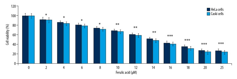
BACKGROUND Ferulic acid is an antioxidant phenolic compound derived from plants, which has effects on cancer cells. This study aimed to investigate the effects of ferulic acid on HeLa and Caski human cervical carcinoma cells and the molecular mechanisms involved. MATERIAL AND METHODS HeLa and Caski human cervical carcinoma cells were grown in culture and treated with increasing doses of ferulic acid. The MTT assay was used to evaluate cell viability. Flow cytometry was performed with 4',6-diamidino-2-phenylindole (DAPI) and Annexin V staining for cell apoptosis. The expression of myeloid leukemia cell differentiation-1 (Mcl-1) protein and MCL-1 mRNA were determined by Western blot and reverse transcription-polymerase chain reaction (RT-PCR). RESULTS Ferulic acid significantly reduced HeLa and Caski cell viability in the concentration range of 4-20 µM (P<0.05). Ferulic acid treatment promoted DNA condensation and significantly increased apoptosis in Caski cells (P<0.05). Ferulic acid treatment resulted in the activation of pro-caspase-3, pro-caspase-8, pro-caspase-9, and PARP. The MTT assay showed that ferulic acid did not reduce the viability of Caski cells treated with the caspase inhibitor, z-VAD-fmk. Ferulic acid reduced the levels of Bcl-2 and Mcl-1, and increased the levels of Bax and reactive oxygen species (ROS). In Caski cells, Akt and PI3K phosphorylation were reduced by ferulic acid in a concentration-dependent manner. CONCLUSIONS The effects of ferulic acid were dose-dependent and resulted in cell cytotoxicity and apoptosis of HeLa and Caski cells, and the PI3K/Akt signaling pathway was down-regulated in Caski cells.
阿魏酸是一种从植物中提取的抗氧化性酚类化合物,对癌细胞有作用。本研究旨在探讨阿魏酸对人宫颈癌细胞 HeLa 和 Caski 的影响及其相关分子机制。
在培养条件下培养 HeLa 和 Caski 人宫颈癌细胞,并给予递增剂量的阿魏酸处理。采用 MTT 法评估细胞活力。用 4',6-二脒基-2-苯基吲哚(DAPI)和 Annexin V 染色进行细胞凋亡的流式细胞术分析。用 Western blot 和逆转录-聚合酶链反应(RT-PCR)测定髓样白血病细胞分化-1(Mcl-1)蛋白和 MCL-1mRNA 的表达。
阿魏酸在 4-20μM 浓度范围内显著降低 HeLa 和 Caski 细胞活力(P<0.05)。阿魏酸处理促进 Caski 细胞中 DNA 凝聚并显著增加细胞凋亡(P<0.05)。阿魏酸处理导致原胱天蛋白酶-3、原胱天蛋白酶-8、原胱天蛋白酶-9 和 PARP 的激活。MTT 试验表明,用胱天蛋白酶抑制剂 z-VAD-fmk 处理 Caski 细胞后,阿魏酸并未降低细胞活力。阿魏酸降低了 Bcl-2 和 Mcl-1 的水平,增加了 Bax 和活性氧(ROS)的水平。在 Caski 细胞中,阿魏酸呈浓度依赖性地降低 Akt 和 PI3K 的磷酸化。
阿魏酸的作用呈剂量依赖性,导致 HeLa 和 Caski 细胞的细胞毒性和细胞凋亡,并且在 Caski 细胞中下调了 PI3K/Akt 信号通路。