Gong Liang, Li Huaisu, Yang Dan, Peng Yinwei, Liu Duan, Zhong Ming, Zhang Bei, Xu Ronghua, Kang Jian
Department of Neurology, Chengdu Second People's Hospital, Chengdu, China.
Hospital of Chengdu University of Traditional Chinese Medicine, Chengdu, China.
Front Neurosci. 2020 Jan 9;13:1353. doi: 10.3389/fnins.2019.01353. eCollection 2019.
Excessive daytime sleepiness (EDS) is one of the common and burdensome non-motor symptoms of Parkinson's disease (PD). However, the underlying neuropathology mechanism in PD patients with EDS (PD-EDS) remains unclear. The present study aims to delineate potential locations of structural alteration of subcortical regions in early stage and drug-naïve PD-EDS.
The study had 252 patients with PD and 92 matched healthy controls (HC). EDS was estimated with the Epworth Sleepiness Scale, with a cutoff of 10. Ultimately, 59 patients were considered as PD-EDS. The remaining 193 were PD patients without EDS (PD-nEDS). FMRIB's Integrated Registration and Segmentation Tool (FIRST) was employed to assess the volumetric and surface alterations of subcortical nuclei in PD and PD-EDS.
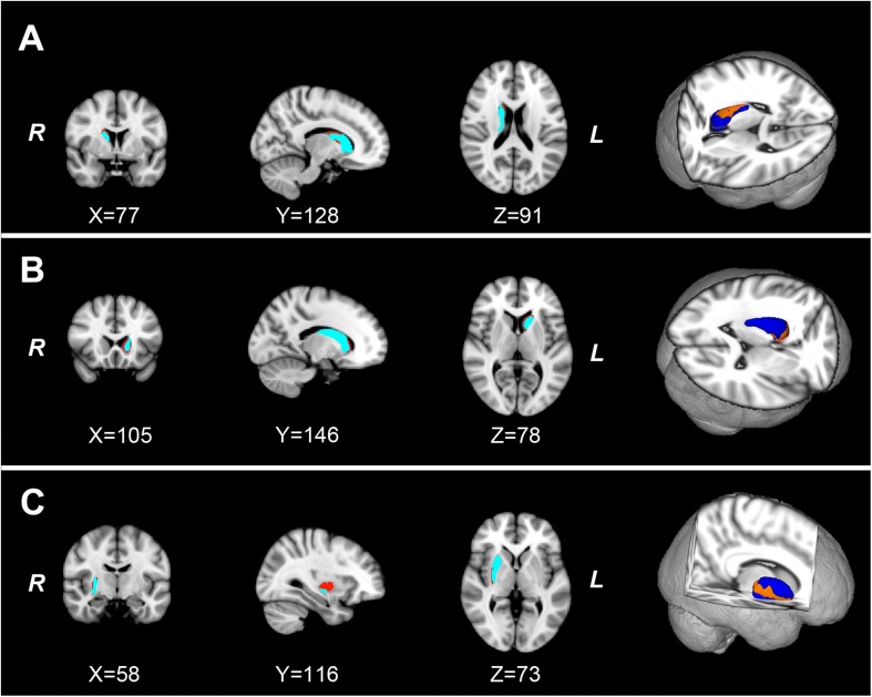
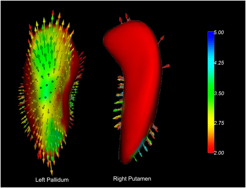
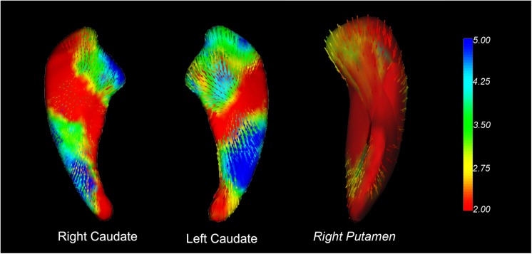
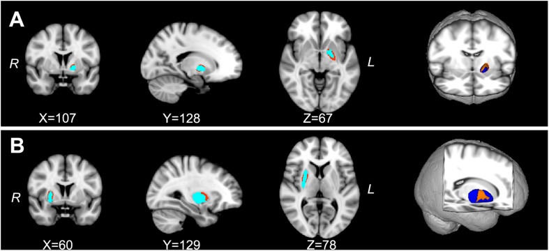
Volumetric analyses found no difference in the subcortical nucleus volume between PD and HC, or PD-EDS and PD-nEDS groups. The shape analyses revealed the local atrophic changes in bilateral caudate and right putamen in patients with PD. In addition, the hypertrophic changes were located in the right putamen and left pallidum in PD-EDS than in PD-nEDS.
Our findings revealed the regional hypertrophy of the striatum in PD-EDS. Our results indicate that local hypertrophic striatum would be a valuable early biomarker for detecting the alteration in PD-EDS. The shape analysis contributes valuable information when investigating PD-EDS.
日间过度嗜睡(EDS)是帕金森病(PD)常见且负担较重的非运动症状之一。然而,伴有EDS的PD患者(PD-EDS)潜在的神经病理学机制仍不清楚。本研究旨在描绘早期未用药的PD-EDS患者皮质下区域结构改变的潜在位置。
该研究纳入了252例PD患者和92例匹配的健康对照(HC)。采用爱泼沃斯嗜睡量表评估EDS,临界值为10分。最终,59例患者被视为PD-EDS。其余193例为无EDS的PD患者(PD-nEDS)。使用FMRIB的综合注册与分割工具(FIRST)评估PD和PD-EDS患者皮质下核团的体积和表面改变。
体积分析发现,PD组与HC组之间、PD-EDS组与PD-nEDS组之间皮质下核团体积无差异。形状分析显示,PD患者双侧尾状核和右侧壳核存在局部萎缩性改变。此外,与PD-nEDS相比,PD-EDS患者右侧壳核和左侧苍白球存在肥厚性改变。
我们的研究结果揭示了PD-EDS患者纹状体区域肥厚。我们的结果表明,局部肥厚的纹状体将是检测PD-EDS改变的有价值的早期生物标志物。在研究PD-EDS时,形状分析提供了有价值的信息。