• 文献检索
  • 文档翻译
  • 深度研究
  • 学术资讯
  • Suppr Zotero 插件Zotero 插件
  • 邀请有礼
  • 套餐&价格
  • 历史记录
应用&插件
Suppr Zotero 插件Zotero 插件浏览器插件Mac 客户端Windows 客户端微信小程序
定价
高级版会员购买积分包购买API积分包
服务
文献检索文档翻译深度研究API 文档MCP 服务
关于我们
关于 Suppr公司介绍联系我们用户协议隐私条款
关注我们

Suppr 超能文献

核心技术专利:CN118964589B侵权必究
粤ICP备2023148730 号-1Suppr @ 2026

文献检索

告别复杂PubMed语法,用中文像聊天一样搜索,搜遍4000万医学文献。AI智能推荐,让科研检索更轻松。

立即免费搜索

文件翻译

保留排版,准确专业,支持PDF/Word/PPT等文件格式,支持 12+语言互译。

免费翻译文档

深度研究

AI帮你快速写综述,25分钟生成高质量综述,智能提取关键信息,辅助科研写作。

立即免费体验

肝硬化和门静脉高压食管静脉曲张患者首次上消化道出血的计算机断层扫描风险评估

Risk assessment of first upper gastrointestinal bleeding using computerized tomoscanning in esophageal varices patients with cirrhosis and portal hypertension.

作者信息

Xie Wei, Chen Fa-Xiang, Zhu Li-Yao, Wen Cheng-Cai, Zhang Xin

机构信息

Department of Radiology, Central Hospital of Wuhan, Tongji Medical College, Huazhong University of Science and Technology, Wuhan.

Department of Hepatology, The Fourth People's Hospital of Huai'an, Jiangsu.

出版信息

Medicine (Baltimore). 2020 Jan;99(5):e18923. doi: 10.1097/MD.0000000000018923.

DOI:10.1097/MD.0000000000018923
PMID:32000404
原文链接:https://pmc.ncbi.nlm.nih.gov/articles/PMC7004787/
Abstract

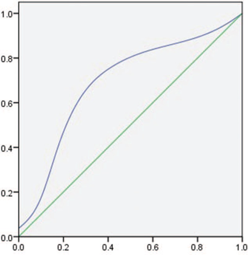
To evaluate the risk of first upper gastrointestinal bleeding by computerized tomoscanning (CT) for esophageal varices patients with cirrhotic portal hypertension.One hundred thirty two esophageal varices patients with cirrhotic portal hypertension who are also complicated with gastrointestinal bleeding were recruited as bleeding group, while another 132 patients without bleeding as non-bleeding group. The diameter of esophageal varices, number of vascular sections, and total area of blood vessels were measured by CT scanning. The sensitivity and specificity of these indicators were calculated, and Youden index was adjusted with the critical point.The diameter of esophageal varices was 7.83 ± 2.76 mm in bleeding group, and 6.57 ± 3.42 mm in non-bleeding group. The Youden index was 0.32 with the critical point 5.55 mm. The area under the receiver operating characteristics (AUROC) was 0.72. The number of venous vessels was 4.5 ± 2 in bleeding group, whereas being 4 ± 2 in non-bleeding group. The Youden index was 0.35 with a critical point 4, and the area under the curve (AUC) was 0.68. The blood vessel area was 1.73 ± 1.15 cm in bleeding group, and 1.12 ± 0.89 cm in non-bleeding group. The Youden index was 0.48 with the critical point being 1.03 cm, and corresponding AUC was 0.82.Among all 3 indicators of the total area, diameter, and number of sections of the esophageal varices, the total area of esophageal varices showed more accuracy as a potential and novel indicator for bleeding prediction.

摘要

评估计算机断层扫描(CT)对肝硬化门静脉高压食管静脉曲张患者首次上消化道出血的风险。选取132例肝硬化门静脉高压合并食管静脉曲张且并发消化道出血的患者作为出血组,另132例未出血患者作为非出血组。通过CT扫描测量食管静脉曲张的直径、血管节段数和血管总面积。计算这些指标的敏感性和特异性,并根据临界点调整约登指数。出血组食管静脉曲张直径为7.83±2.76mm,非出血组为6.57±3.42mm。临界点为5.55mm时,约登指数为0.32。受试者工作特征曲线下面积(AUROC)为0.72。出血组静脉血管数为4.5±2,非出血组为4±2。临界点为4时,约登指数为0.35,曲线下面积(AUC)为0.68。出血组血管面积为1.73±1.15cm,非出血组为1.12±0.89cm。临界点为1.03cm时,约登指数为0.48,相应的AUC为0.82。在食管静脉曲张的总面积、直径和节段数这3个指标中,食管静脉曲张总面积作为出血预测的潜在新指标显示出更高的准确性。

https://cdn.ncbi.nlm.nih.gov/pmc/blobs/d3dc/7004787/991254210230/medi-99-e18923-g010.jpg
https://cdn.ncbi.nlm.nih.gov/pmc/blobs/d3dc/7004787/86ecceb0825b/medi-99-e18923-g001.jpg
https://cdn.ncbi.nlm.nih.gov/pmc/blobs/d3dc/7004787/a0ceaad12443/medi-99-e18923-g002.jpg
https://cdn.ncbi.nlm.nih.gov/pmc/blobs/d3dc/7004787/e247d7679616/medi-99-e18923-g003.jpg
https://cdn.ncbi.nlm.nih.gov/pmc/blobs/d3dc/7004787/a7fc4b58cb3d/medi-99-e18923-g004.jpg
https://cdn.ncbi.nlm.nih.gov/pmc/blobs/d3dc/7004787/e105b537079c/medi-99-e18923-g005.jpg
https://cdn.ncbi.nlm.nih.gov/pmc/blobs/d3dc/7004787/b98ee1d07b25/medi-99-e18923-g006.jpg
https://cdn.ncbi.nlm.nih.gov/pmc/blobs/d3dc/7004787/c9e82ab2f367/medi-99-e18923-g008.jpg
https://cdn.ncbi.nlm.nih.gov/pmc/blobs/d3dc/7004787/3a707396f4c0/medi-99-e18923-g009.jpg
https://cdn.ncbi.nlm.nih.gov/pmc/blobs/d3dc/7004787/991254210230/medi-99-e18923-g010.jpg

相似文献

1
Risk assessment of first upper gastrointestinal bleeding using computerized tomoscanning in esophageal varices patients with cirrhosis and portal hypertension.肝硬化和门静脉高压食管静脉曲张患者首次上消化道出血的计算机断层扫描风险评估
Medicine (Baltimore). 2020 Jan;99(5):e18923. doi: 10.1097/MD.0000000000018923.
2
Non-variceal upper gastrointestinal bleeding in cirrhotic patients in Nile Delta.尼罗河三角洲肝硬化患者的非静脉曲张性上消化道出血
Indian J Gastroenterol. 2016 Jan;35(1):25-32. doi: 10.1007/s12664-016-0622-7. Epub 2016 Feb 17.
3
Hemodynamic analysis of esophageal varices in patients with liver cirrhosis using color Doppler ultrasound.利用彩色多普勒超声对肝硬化患者食管静脉曲张进行血流动力学分析。
World J Gastroenterol. 2005 Aug 7;11(29):4560-5. doi: 10.3748/wjg.v11.i29.4560.
4
[New possibilities of Doppler flowmetric ultrasound diagnosis of portal hypertension complicated with varicose veins of the esophagus and bleeding prediction].[多普勒血流超声诊断门静脉高压合并食管静脉曲张及出血预测的新可能性]
Lik Sprava. 2013 Dec(8):74-9.
5
Risk of esophageal variceal bleeding based on endoscopic ultrasound evaluation of the sum of esophageal variceal cross-sectional surface area.基于内镜超声评估食管静脉曲张横截面积总和的食管静脉曲张出血风险
Am J Gastroenterol. 2003 Feb;98(2):454-9. doi: 10.1111/j.1572-0241.2003.07224.x.
6
Clinical and laboratory predictors of esophageal varices in children and adolescents with portal hypertension syndrome.门静脉高压综合征儿童及青少年食管静脉曲张的临床及实验室预测指标
J Pediatr Gastroenterol Nutr. 2008 Feb;46(2):178-83. doi: 10.1097/MPG.0b013e318156ff07.
7
Role of Serum Soluble CD163 in the Diagnosis, Risk of Bleeding, and Prognosis of Gastro-Esophageal Varices in Cirrhotic Patients.血清可溶性CD163在肝硬化患者胃食管静脉曲张诊断、出血风险及预后中的作用
J Interferon Cytokine Res. 2017 Mar;37(3):112-118. doi: 10.1089/jir.2016.0098.
8
[Correlation of Doppler ultrasound of the portal system with endoscopic changes caused by portal hypertension in cirrhotic patients].[肝硬化患者门静脉系统多普勒超声与门静脉高压所致内镜改变的相关性]
Arq Gastroenterol. 2006 Jul-Sep;43(3):178-83. doi: 10.1590/s0004-28032006000300005.
9
[Color duplex Doppler ultrasonography in the evaluation of the risk of esophageal varices bleeding in cirrhotic patients].[彩色双功多普勒超声评估肝硬化患者食管静脉曲张出血风险]
Gastroenterol Hepatol. 2000 Dec;23(10):466-9.
10
Transient Elastography Identifies the Risk of Esophageal Varices and Bleeding in Patients With Hepatitis B Virus-Related Liver Cirrhosis.瞬时弹性成像可识别乙型肝炎病毒相关性肝硬化患者食管静脉曲张及出血风险。
Ultrasound Q. 2018 Sep;34(3):141-147. doi: 10.1097/RUQ.0000000000000373.

引用本文的文献

1
The role of computed tomography for the prediction of esophageal variceal bleeding: Current status and future perspectives.计算机断层扫描在预测食管静脉曲张出血中的作用:现状与未来展望。
World J Gastrointest Endosc. 2023 Dec 16;15(12):681-689. doi: 10.4253/wjge.v15.i12.681.
2
Assessing the Predictive Factors for Bleeding in Esophageal Variceal Disease: A Systematic Review.评估食管静脉曲张疾病出血的预测因素:一项系统评价。
Cureus. 2023 Nov 17;15(11):e48954. doi: 10.7759/cureus.48954. eCollection 2023 Nov.

本文引用的文献

1
Management Options for Gastric Variceal Hemorrhage.胃静脉曲张出血的处理方法。
JAMA Surg. 2019 Jun 1;154(6):540-548. doi: 10.1001/jamasurg.2019.0407.
2
Predictive value of CT for first esophageal variceal bleeding in patients with cirrhosis: Score calibration and prediction.CT对肝硬化患者首次食管静脉曲张出血的预测价值:评分校准与预测
Eur J Radiol. 2017 Aug;93:289. doi: 10.1016/j.ejrad.2017.05.009. Epub 2017 May 9.
3
Bleeding esophageal varices: Who should receive a shunt?出血性食管静脉曲张:谁应该接受分流术?
Cleve Clin J Med. 2017 Mar;84(3):199-201. doi: 10.3949/ccjm.84a.15149.
4
Predictive value of CT for first esophageal variceal bleeding in patients with cirrhosis: Value of para-umbilical vein patency.CT对肝硬化患者首次食管静脉曲张出血的预测价值:脐旁静脉通畅情况的价值
Eur J Radiol. 2017 Feb;87:45-52. doi: 10.1016/j.ejrad.2016.12.006. Epub 2016 Dec 9.
5
Portal hypertensive bleeding in cirrhosis: Risk stratification, diagnosis, and management: 2016 practice guidance by the American Association for the study of liver diseases.肝硬化门静脉高压出血:风险分层、诊断及管理:美国肝病研究协会2016年实践指南
Hepatology. 2017 Jan;65(1):310-335. doi: 10.1002/hep.28906. Epub 2016 Dec 1.
6
A novel model to predict esophageal varices in patients with compensated cirrhosis using acoustic radiation force impulse elastography.一种使用声辐射力脉冲弹性成像预测代偿期肝硬化患者食管静脉曲张的新模型。
PLoS One. 2015 Mar 31;10(3):e0121009. doi: 10.1371/journal.pone.0121009. eCollection 2015.
7
Endoscopic treatment of esophageal varices in patients with liver cirrhosis.肝硬化患者食管静脉曲张的内镜治疗
World J Gastroenterol. 2014 Sep 28;20(36):13015-26. doi: 10.3748/wjg.v20.i36.13015.
8
Prevention of portal hypertension: from variceal development to clinical decompensation.门静脉高压的预防:从静脉曲张形成到临床失代偿
Hepatology. 2015 Jan;61(1):375-81. doi: 10.1002/hep.27249. Epub 2014 Sep 26.
9
Clinically severe portal hypertension: role of multi-detector row CT features in diagnosis.临床严重门静脉高压症:多排 CT 特征在诊断中的作用。
Dig Dis Sci. 2014 Sep;59(9):2333-43. doi: 10.1007/s10620-014-3149-8. Epub 2014 Apr 11.
10
Invasive and non-invasive techniques for detecting portal hypertension and predicting variceal bleeding in cirrhosis: a review.肝硬化门静脉高压症及静脉曲张出血的侵袭性和非侵袭性检测技术:综述。
Ann Med. 2014 Feb;46(1):8-17. doi: 10.3109/07853890.2013.857831. Epub 2013 Dec 16.