Department of Respiratory Medicine, Saitama Red Cross Hospital, 1-5 Shintoshin, Chuo-ku, Saitama, 330-8553, Japan.
Department of Internal Medicine, Division of Respiratory Medicine, Jikei University School of Medicine, Tokyo, Japan.
BMC Pulm Med. 2020 Jan 30;20(1):25. doi: 10.1186/s12890-020-1061-x.
Interstitial lung disease (ILD) is the most common and important pulmonary manifestation of rheumatoid arthritis (RA). A radiological honeycomb pattern has been described in diverse forms of ILD that can impact survival. However, the clinical course and sequential radiological changes in the formation of the honeycomb pattern in patients with RA-ILD is not fully understood.
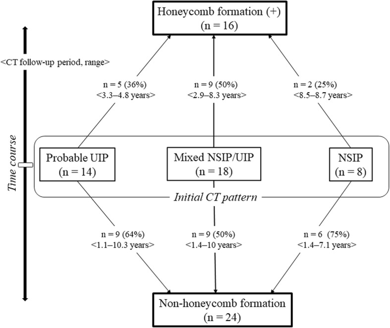
We evaluated the sequential changes in computed tomography findings in 40 patients with chronic forms of RA-ILD without the honeycomb pattern at initial diagnosis. We classified the patients into the Non-honeycomb group and Honeycomb group, and then analyzed the characteristics and prognosis of the two groups. The term "honeycomb formation" indicated a positive finding of honeycombing on any available follow-up CT.
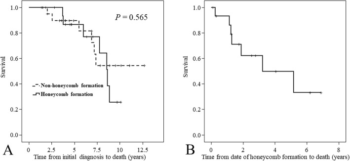
Our RA-ILD cohort included patients with probable usual interstitial pneumonia (UIP) (35%), nonspecific interstitial pneumonia (NSIP) (20%), and mixed NSIP/UIP (45%). Among all RA-ILD patients, 16 (40%) showed honeycomb formation on follow-up CT (median time between initial and last follow-up CT was 4.7 years). Patient characteristics and prognosis were not significantly different between the Non-honeycomb and Honeycomb groups. However, Kaplan-Meier survival curve for the time from the date of honeycomb formation to death showed a poor median survival time of 3.2 years.
A certain number of patients with RA-ILD developed a honeycomb pattern during long-term follow-up, regardless of whether they had UIP or NSIP. Prognosis in the patients with characteristics of both progressive ILD and honeycomb formation could be poor. Although radiological findings over the disease course and clinical disease behavior in RA-ILD are heterogenous, clinicians should be alert to the possibility of progressive disease and poor prognosis in patients with RA-ILD who form a honeycomb pattern during follow-up observation.
间质性肺疾病(ILD)是类风湿关节炎(RA)最常见和最重要的肺部表现。在不同形式的ILD 中已经描述了一种影像学蜂窝状模式,它会影响生存。然而,RA-ILD 患者中蜂窝状模式形成的临床病程和连续影像学变化尚不完全清楚。
我们评估了 40 例慢性 RA-ILD 患者在初始诊断时无蜂窝状模式的 CT 检查结果的连续变化。我们将患者分为非蜂窝组和蜂窝组,然后分析两组的特征和预后。“蜂窝形成”一词表示在任何可获得的随访 CT 上存在蜂窝的阳性发现。
我们的 RA-ILD 队列包括可能的寻常型间质性肺炎(UIP)(35%)、非特异性间质性肺炎(NSIP)(20%)和混合 NSIP/UIP(45%)患者。在所有 RA-ILD 患者中,16 例(40%)在随访 CT 上显示蜂窝形成(初始和最后一次随访 CT 之间的中位时间为 4.7 年)。非蜂窝组和蜂窝组患者的特征和预后无显著差异。然而,从出现蜂窝的日期到死亡的时间的 Kaplan-Meier 生存曲线显示,中位生存时间为 3.2 年,预后较差。
一定数量的 RA-ILD 患者在长期随访中出现蜂窝状模式,无论其是否存在 UIP 或 NSIP。具有进行性ILD 和蜂窝形成特征的患者的预后可能较差。尽管 RA-ILD 的病程中的影像学表现和临床疾病行为具有异质性,但临床医生应该警惕在随访观察期间出现蜂窝状模式的 RA-ILD 患者疾病进展和预后不良的可能性。