Johansson Junko, Kiffin Roberta, Aydin Ebru, Nilsson Malin S, Hellstrand Kristoffer, Lindnér Per, Naredi Peter, Olofsson Bagge Roger, Martner Anna
TIMM Laboratory, Sahlgrenska Cancer Center, Sahlgrenska Academy, University of Gothenburg, Gothenburg, Sweden.
Department of Surgery, Institute of Clinical Sciences, Sahlgrenska Academy, University of Gothenburg, Gothenburg, Sweden.
Oncoimmunology. 2019 Nov 3;9(1):1684126. doi: 10.1080/2162402X.2019.1684126. eCollection 2020.
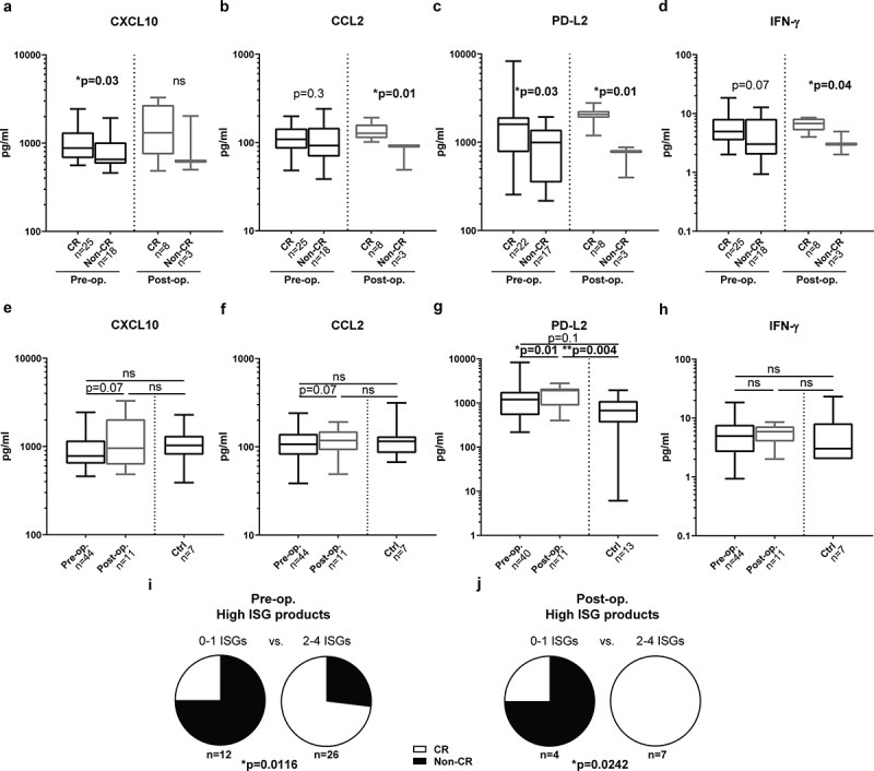
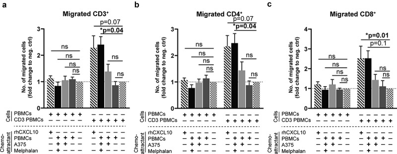
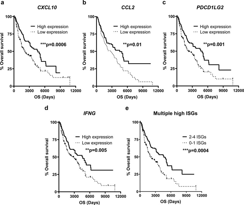
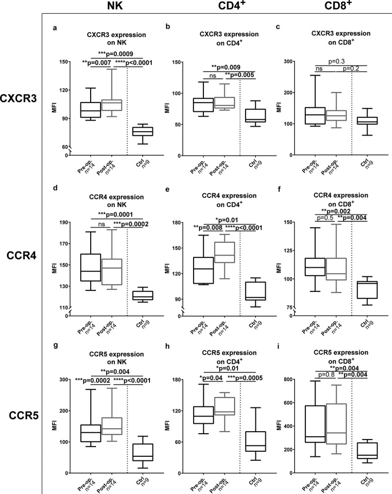
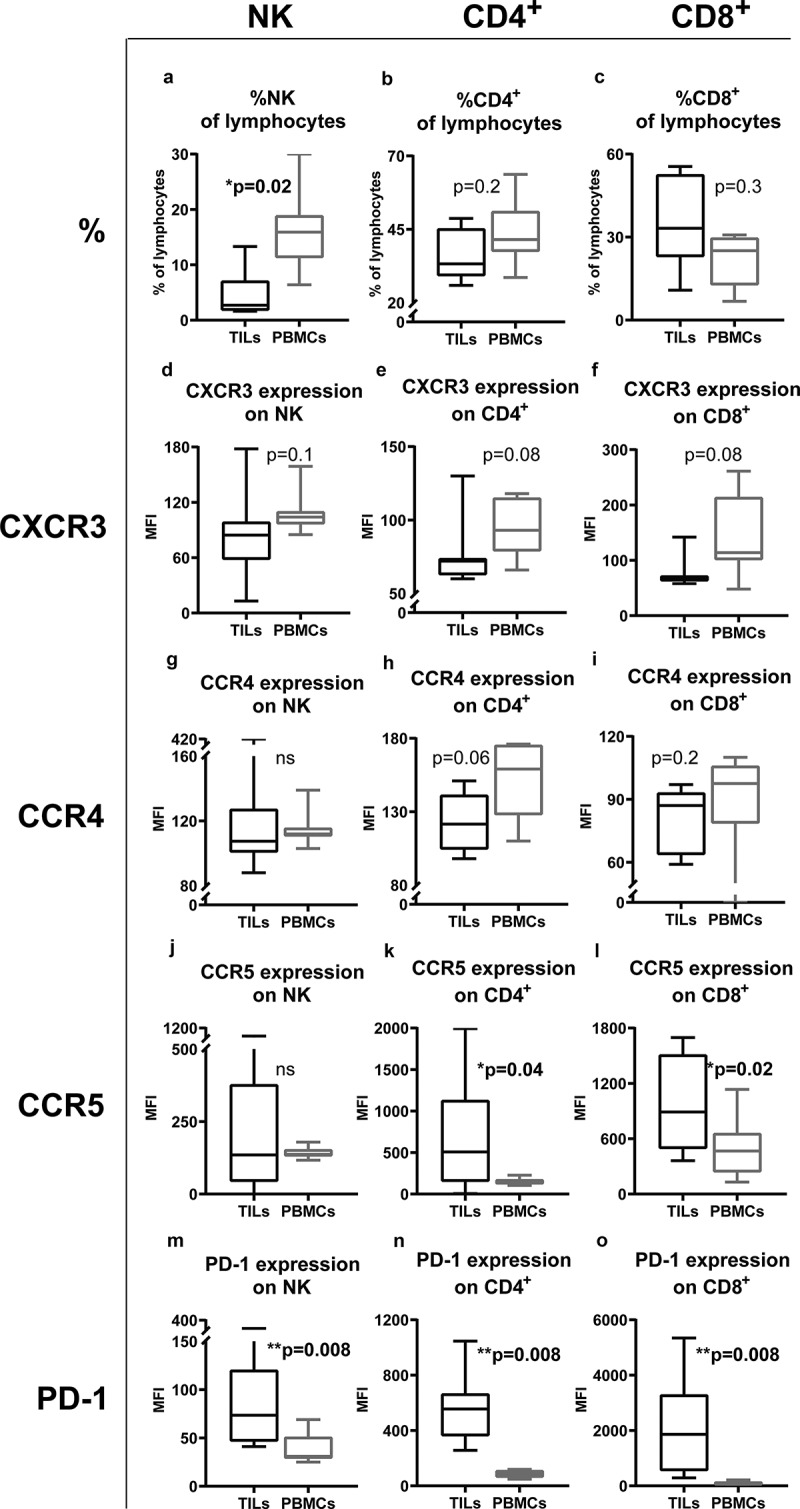
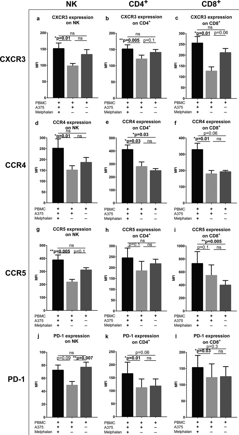
Hyperthermic isolated limb perfusion (ILP) with high-dose melphalan is a treatment option for melanoma patients with metastasis confined to limbs (in-transit metastasis). The therapy entails a complete response (CR) rate of 50-70%. Cellular immunity is proposed to impact on the clinical efficacy of ILP, but the detailed aspects of ILP-induced immune activation remain to be explored. For this study, we explored the potential role of interferon-stimulated gene (ISG) products, including CXCL10, CCL2, PD-L2 and IFN-γ along with expression of their cognate receptors CXCR3, CCR4, CCR5 and PD-1 on lymphocytes, for the clinical efficacy of ILP. Patients with high serum levels of CXCL10, CCL2, PD-L2 and IFN-γ were more likely to achieve CR after ILP. Additionally, the expression of CXCR3, CCR4 and CCR5 on T cells and/or natural killer (NK) cells was enhanced by ILP. Peripheral blood mononuclear cells (PBMCs) secreted high levels of CXCL10, CCL2 and IFN-γ in response to co-culture with melphalan-exposed melanoma cells . Activated T cells migrated toward supernatants from these co-cultures. Furthermore, melphalan-exposed melanoma cells triggered upregulation of CXCR3, CCR4, CCR5 and PD-1 on co-cultured T cells and/or NK cells. Our results suggest that constituents released from melphalan-exposed melanoma cells stimulate the ISG axis with ensuing formation of chemokines and upregulation of chemokine receptor expression on anti-neoplastic immune cells, which may contribute in ILP-induced tumor regression.
高剂量美法仑热灌注隔离肢体(ILP)是治疗转移局限于肢体(移行转移)的黑色素瘤患者的一种治疗选择。该疗法的完全缓解(CR)率为50-70%。细胞免疫被认为会影响ILP的临床疗效,但ILP诱导的免疫激活的详细情况仍有待探索。在本研究中,我们探讨了干扰素刺激基因(ISG)产物,包括CXCL10、CCL2、PD-L2和IFN-γ及其在淋巴细胞上的同源受体CXCR3、CCR4、CCR5和PD-1的表达对ILP临床疗效的潜在作用。血清中CXCL10、CCL2、PD-L2和IFN-γ水平高的患者在接受ILP治疗后更有可能实现CR。此外,ILP可增强T细胞和/或自然杀伤(NK)细胞上CXCR3、CCR4和CCR5的表达。外周血单个核细胞(PBMC)在与暴露于美法仑的黑色素瘤细胞共培养时分泌高水平的CXCL10、CCL2和IFN-γ。活化的T细胞向这些共培养物的上清液迁移。此外,暴露于美法仑的黑色素瘤细胞可触发共培养的T细胞和/或NK细胞上CXCR3、CCR4、CCR5和PD-1的上调。我们的结果表明,暴露于美法仑的黑色素瘤细胞释放的成分刺激ISG轴,随后形成趋化因子并上调抗肿瘤免疫细胞上趋化因子受体的表达,这可能有助于ILP诱导的肿瘤消退。