Discipline of Pathology, Sydney Medical School, Brain and Mind Centre, The University of Sydney, Sydney, New South Wales, Australia.
Department of Neuropathology, Royal Prince Alfred Hospital, Sydney, New South Wales, Australia.
PLoS One. 2020 Jan 31;15(1):e0228226. doi: 10.1371/journal.pone.0228226. eCollection 2020.
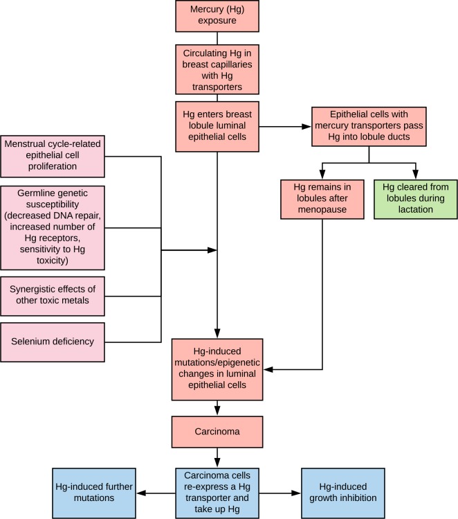
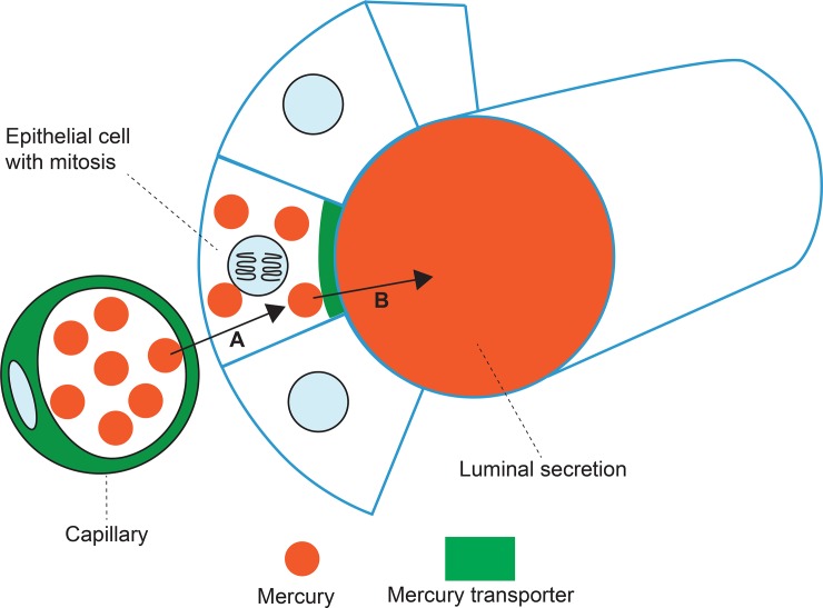
Exposure to toxic metals such as mercury has been proposed to be a risk factor for the development of breast cancer since some metals can promote genetic mutations and epigenetic changes. We sought to find what toxic metals are present in normal breast tissue and in the tumours of women who had mastectomies for invasive ductal breast carcinoma.
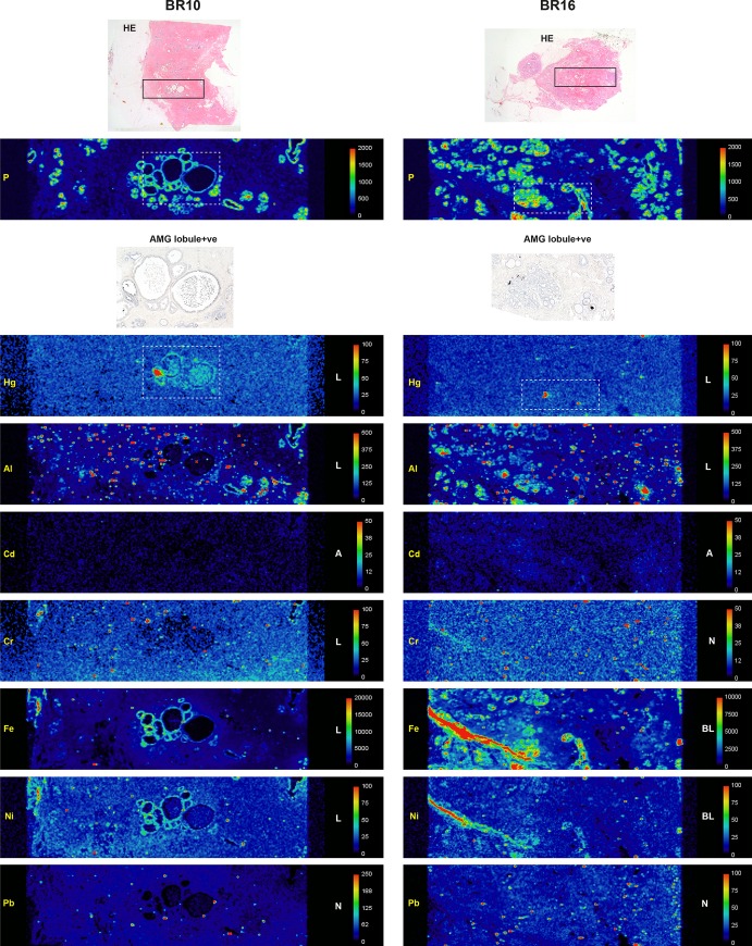
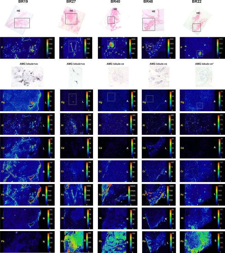
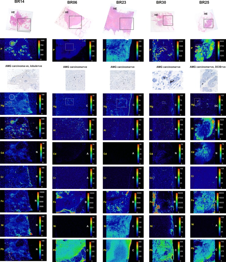
Formalin-fixed paraffin-embedded blocks from mastectomies for breast carcinoma were examined from 50 women aged 34-69 years. Paraffin blocks selected for elemental analysis were from breast tissue not involved by carcinoma and from the carcinoma itself. Seven micrometer-thick sections were stained with autometallography to demonstrate the presence of mercury, and subjected to laser ablation-inductively coupled plasma-mass spectrometry (LA-ICP-MS) to confirm the presence of mercury and to detect other toxic metals.
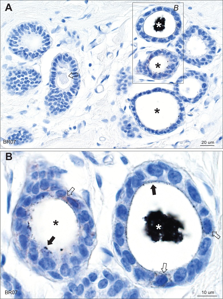
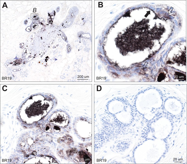
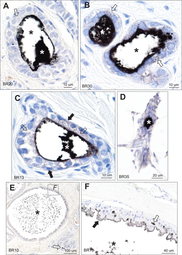
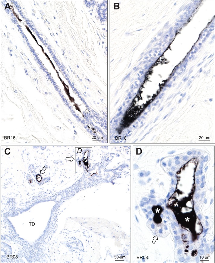
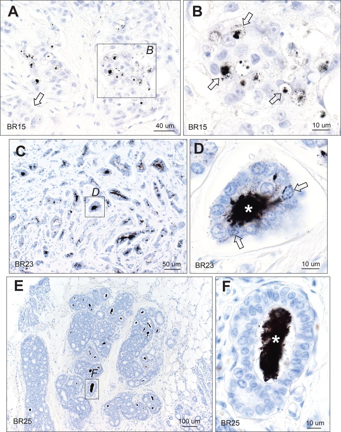
Autometallography-detected mercury was seen in intraductal secretions and some luminal epithelial cells of normal breast lobules in 26 (55%) of the 47 samples where lobules were present, and in 10 (23%) of carcinomas from the 44 samples where carcinoma was present. In eight samples ductal carcinoma in situ was present and one of these contained mercury. LA-ICP-MS confirmed the presence of mercury in samples that stained with autometallography, and detected lead, iron, nickel, aluminium, chromium and cadmium in some samples.
Mercury was present in normal breast lobules in more than half of mastectomy samples that contained an invasive carcinoma, and in a smaller proportion of carcinomas and ductal carcinomas in situ. Other toxic metals that may interact synergistically with mercury could be detected in some samples. These findings do not provide direct evidence that toxic metals such as mercury play a role in the pathogenesis of breast cancer, but suggest that future molecular biological investigations on the role of toxic metals in breast cancer are warranted.
由于一些金属可以促进基因突变和表观遗传改变,因此接触有毒金属(如汞)被认为是乳腺癌发展的一个危险因素。我们试图寻找在接受乳房切除术治疗浸润性导管乳腺癌的女性的正常乳房组织和肿瘤中存在哪些有毒金属。
对 50 名年龄在 34-69 岁的女性乳房切除术的福尔马林固定石蜡包埋块进行了检查。选择进行元素分析的石蜡块来自未受癌影响的乳房组织和癌本身。对 7 微米厚的切片进行自动金属成像以显示汞的存在,并进行激光烧蚀-电感耦合等离子体质谱(LA-ICP-MS)以确认汞的存在并检测其他有毒金属。
在 47 个存在小叶的样本中的 26 个(55%)和在 44 个存在癌的样本中的 10 个(23%)中,可见自动金属成像检测到的汞存在于正常小叶的管内分泌物和一些腔上皮细胞中。在 8 个样本中存在导管原位癌,其中一个样本含有汞。LA-ICP-MS 证实了自动金属成像染色样本中汞的存在,并在一些样本中检测到铅、铁、镍、铝、铬和镉。
在含有浸润性癌的乳房切除术样本中,超过一半的正常小叶中存在汞,在较小比例的癌和导管原位癌中也存在汞。在一些样本中还可以检测到其他可能与汞协同作用的有毒金属。这些发现并没有直接证据表明有毒金属(如汞)在乳腺癌的发病机制中起作用,但表明有必要对有毒金属在乳腺癌中的作用进行未来的分子生物学研究。