Department of Oral and Maxillofacial Surgery, School of Dentistry, Shiraz University of Medical Sciences, Shiraz, Iran.
BMC Oral Health. 2020 Jan 31;20(1):31. doi: 10.1186/s12903-020-1019-7.
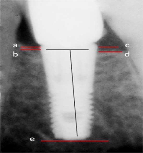
To evaluated the marginal bone loss around dental implants by two insertion methods.
Eligible patients were divided into two groups; manual and mechanized groups. Peri-apical x-ray using a customized device to standardize the radiographs designed and used to take three periodical radiographs; after surgery, three months, and six months follow up. An independent t-test was used to compare the two groups regarding the average level of marginal bone loss (p < 0.05).
After excluding dropouts, a total of 273 patients (120 males and 153 females, aged between 25 and 67 years old) were included in the study. The average marginal bone loss in the manual insertion method was 0.44 ± 0.84 mm, and 0.59 ± 0.20 mm, and for the mechanized method was 0.51 ± 0.20 mm and 0.67 ± 0.19 mm after three and six months, respectively. There was a significant difference in marginal bone loss after six months between the two groups(p < 0.001). However, no differences were observed after three months (p = 0.24).
Under the condition of this study, both techniques were safe and resulted in an acceptable amount of bone resorption; however, in the manual method, the less marginal bone loss occurred after six months.
评估两种种植体植入方法的边缘骨丧失情况。
将符合条件的患者分为两组:手动组和机械组。使用专门设计的定制设备进行根尖周 X 光检查,以标准化放射照片,并用于拍摄三个定期放射照片;在手术后、三个月和六个月进行随访。使用独立 t 检验比较两组的平均边缘骨丧失水平(p<0.05)。
排除脱落者后,共有 273 名患者(120 名男性和 153 名女性,年龄在 25 至 67 岁之间)纳入研究。手动植入方法的平均边缘骨丧失量分别为 0.44±0.84mm 和 0.59±0.20mm,机械植入方法分别为 0.51±0.20mm 和 0.67±0.19mm,在三个月和六个月后。两组之间在六个月后的边缘骨丧失量存在显著差异(p<0.001)。然而,在三个月时未观察到差异(p=0.24)。
在本研究条件下,两种技术均安全且导致可接受的骨吸收量;然而,在手动方法中,六个月后边缘骨丧失量较少。