George Washington University and Children's National Hospital, Washington, DC.
Children's National Hospital, Washington, DC.
Arthritis Rheumatol. 2020 Jul;72(7):1170-1183. doi: 10.1002/art.41215. Epub 2020 May 31.
Muscle inflammation is a feature in myositis and Duchenne muscular dystrophy (DMD). Autoimmune mechanisms are thought to contribute to muscle weakness in patients with myositis. However, a lack of correlation between the extent of inflammatory cell infiltration and muscle weakness indicates that nonimmune pathologic mechanisms may play a role. The present study focused on 2 microRNA (miRNA) sets previously identified as being elevated in the muscle of patients with DMD-an "inflammatory" miRNA set that is dampened with glucocorticoids, and a "dystrophin-targeting" miRNA set that inhibits dystrophin translation-to test the hypothesis that these miRNAs are similarly dysregulated in the muscle of patients with myositis, and could contribute to muscle weakness and disease severity.
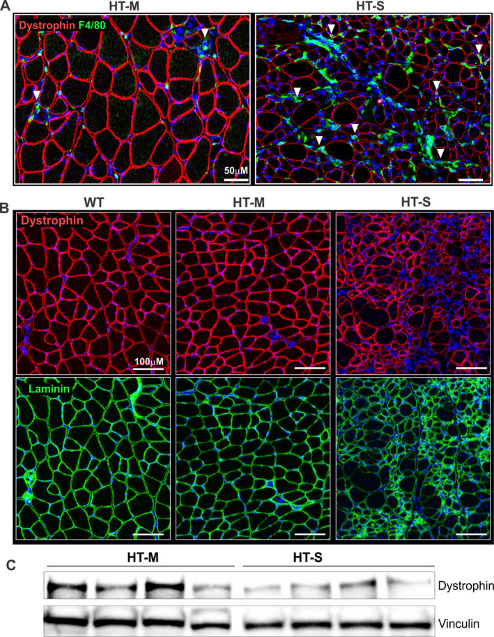
A major histocompatibility complex class I-transgenic mouse model of myositis was utilized to study gene and miRNA expression and histologic features in the muscle tissue, with the findings validated in human muscle biopsy tissue from 6 patients with myositis. Mice were classified as having mild or severe myositis based on transgene expression, body weight, histologic disease severity, and muscle strength/weakness.
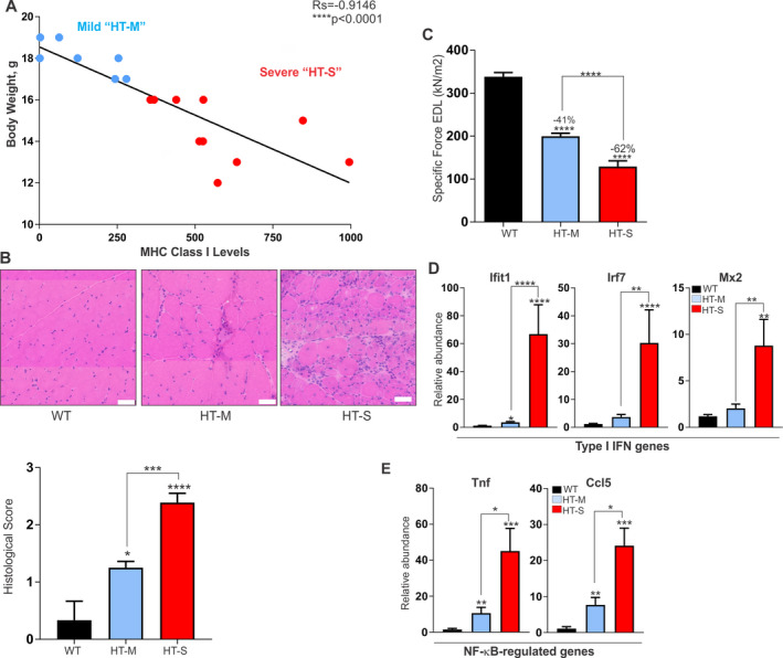
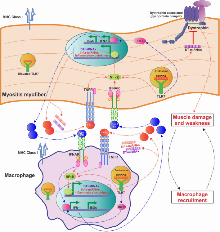
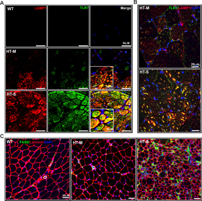
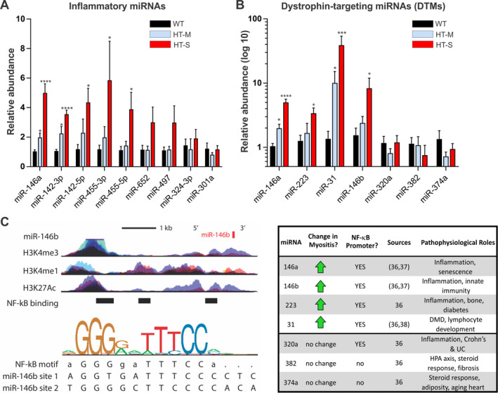
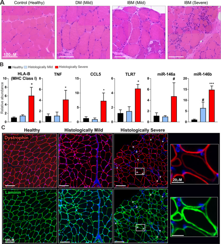
In mice with severe myositis, muscle tissue showed mononuclear cell infiltration along with elevated expression of type I interferon and NF-κB-regulated genes, including Tlr7 (3.8-fold increase, P < 0.05). Furthermore, mice with severe myositis showed elevated expression of inflammatory miRNAs (miR-146a, miR-142-3p, miR-142-5p, miR-455-3p, and miR-455-5p; ~3-40-fold increase, P < 0.05) and dystrophin-targeting miRNAs (miR-146a, miR-146b, miR-31, and miR-223; ~3-38-fold increase, P < 0.05). Bioinformatics analyses of chromatin immunoprecipitation sequencing (ChIP-seq) data identified at least one NF-κB consensus element within the promoter/enhancer regions of these miRNAs. Western blotting and immunofluorescence analyses of the muscle tissue from mice with severe myositis demonstrated reduced levels of dystrophin. In addition, elevated levels of NF-κB-regulated genes, TLR7, and miRNAs along with reduced dystrophin levels were observed in muscle biopsy tissue from patients with histologically severe myositis.
These data demonstrate that an acquired dystrophin deficiency may occur through NF-κB-regulated miRNAs in myositis, thereby suggesting a unifying theme in which muscle injury, inflammation, and weakness are perpetuated both in myositis and in DMD.
肌肉炎症是肌炎和杜氏肌营养不良症(DMD)的特征。自身免疫机制被认为导致肌炎患者的肌肉无力。然而,炎症细胞浸润的程度与肌肉无力之间缺乏相关性表明,非免疫病理机制可能起作用。本研究集中于先前在 DMD 患者肌肉中发现的两组微 RNA(miRNA)-一组被糖皮质激素抑制的“炎症”miRNA 集,以及一组抑制肌营养不良蛋白翻译的“肌营养不良蛋白靶向”miRNA 集-以检验以下假设,即这些 miRNA 在肌炎患者的肌肉中同样失调,并可能导致肌肉无力和疾病严重程度。
利用主要组织相容性复合物 I 转基因小鼠肌炎模型研究肌肉组织中的基因和 miRNA 表达及组织学特征,并在 6 例肌炎患者的肌肉活检组织中验证发现。根据转基因表达、体重、组织学疾病严重程度和肌肉力量/无力,将小鼠分类为轻度或重度肌炎。
在严重肌炎的小鼠中,肌肉组织表现出单核细胞浸润,同时 I 型干扰素和 NF-κB 调节基因的表达升高,包括 Tlr7(增加 3.8 倍,P < 0.05)。此外,严重肌炎的小鼠表现出炎症 miRNA(miR-146a、miR-142-3p、miR-142-5p、miR-455-3p 和 miR-455-5p;增加约 3-40 倍,P < 0.05)和肌营养不良蛋白靶向 miRNA(miR-146a、miR-146b、miR-31 和 miR-223;增加约 3-38 倍,P < 0.05)的表达升高。染色质免疫沉淀测序(ChIP-seq)数据的生物信息学分析确定了这些 miRNA 的启动子/增强子区域内至少一个 NF-κB 共有元件。严重肌炎小鼠肌肉组织的 Western blot 和免疫荧光分析显示肌营养不良蛋白水平降低。此外,在组织学严重肌炎患者的肌肉活检组织中观察到 NF-κB 调节基因、TLR7 和 miRNA 的水平升高以及肌营养不良蛋白水平降低。
这些数据表明,肌炎中 NF-κB 调节的 miRNA 可能导致获得性肌营养不良蛋白缺乏,从而提示在肌炎和 DMD 中,肌肉损伤、炎症和无力均得以持续存在的统一主题。