Aguilera-Correa John Jairo, Garcia-Casas Amaya, Mediero Aranzazu, Romera David, Mulero Francisca, Cuevas-López Irene, Jiménez-Morales Antonia, Esteban Jaime
Clinical Microbiology Department, IIS-Fundacion Jimenez Diaz, UAM, Madrid, Spain.
Department of Materials Science and Engineering, University Carlos III of Madrid, Madrid, Spain.
Front Microbiol. 2020 Jan 17;10:2935. doi: 10.3389/fmicb.2019.02935. eCollection 2019.
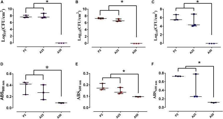
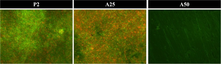
The aim of this study was to evaluate the effect of a moxifloxacin-loaded organic-inorganic sol-gel with different antibiotic concentration in the biofilm development and treatment against , , and , cytotoxicity and cell proliferation of MC3T3-E1 osteoblasts; and its efficacy in preventing the prosthetic joint infection (PJI) caused by clinical strains of and using an murine model. Three bacterial strains, ATCC 35984, 15981, and, ATCC 25922, were used for microbiological studies. Biofilm formation was induced using tryptic-soy supplemented with glucose for 24 h, and then, adhered and planktonic bacteria were estimated using drop plate method and absorbance, respectively. A 24-h-mature biofilm of each species growth in a 96-well plate was treated for 24 h using a MBECTM biofilm Incubator lid with pegs coated with the different types of sol-gel, after incubation, biofilm viability was estimated using alamrBlue. MC3T3-E1 cellular cytotoxicity and proliferation were evaluated using CytoTox 96 Non-Radioactive Cytotoxicity Assay and alamarBlue, respectively. The microbiological studies showed that sol-gel coatings inhibited the biofilm development and treated to a mature biofilm of three evaluated bacterial species. The cell studies showed that the sol-gel both with and without moxifloxacin were non-cytotoxic and that cell proliferation was inversely proportional to the antibiotic concentration containing by sol-gel. In the study, mice weight increased over time, except in the -infected group without coating. The most frequent symptoms associated with infection were limping and piloerection; these symptoms were more frequent in infected groups with non-coated implants than infected groups with coated implants. The response of moxifloxacin-loaded sol-gel to infection was either total or completely absent. No differences in bone mineral density were observed between groups with coated and non-coated implants and macrophage presence lightly increased in the bone grown directly in contact with the antibiotic-loaded sol-gel. In conclusion, moxifloxacin-loaded sol-gel coating is capable of preventing PJI caused by both Gram-positive and Gram-negative species.
本研究旨在评估不同抗生素浓度的载莫西沙星有机-无机溶胶-凝胶对生物膜形成和治疗、耐甲氧西林金黄色葡萄球菌(MRSA)、耐甲氧西林表皮葡萄球菌(MRSE)和耐万古霉素肠球菌(VRE)的效果,MC3T3-E1成骨细胞的细胞毒性和细胞增殖;以及使用小鼠模型预防由MRSA和VRE临床菌株引起的人工关节感染(PJI)的疗效。使用三种细菌菌株,金黄色葡萄球菌ATCC 35984、表皮葡萄球菌15981和粪肠球菌ATCC 25922进行微生物学研究。使用补充有葡萄糖的胰蛋白胨大豆诱导生物膜形成24小时,然后分别使用平板法和吸光度估计粘附菌和浮游菌。在96孔板中生长的每种细菌的24小时成熟生物膜使用带有涂有不同类型溶胶-凝胶的 peg 的MBECTM生物膜培养箱盖处理24小时,孵育后,使用alamrBlue估计生物膜活力。分别使用CytoTox 96非放射性细胞毒性测定法和alamrBlue评估MC3T3-E1细胞的细胞毒性和增殖。微生物学研究表明,溶胶-凝胶涂层抑制生物膜形成并对三种评估细菌物种的成熟生物膜进行处理。细胞研究表明,含莫西沙星和不含莫西沙星的溶胶-凝胶均无细胞毒性,并且细胞增殖与溶胶-凝胶所含抗生素浓度成反比。在小鼠研究中,除了未涂层的VRE感染组外小鼠体重随时间增加。与感染相关最常见的症状是跛行和竖毛;这些症状在未涂层植入物的感染组中比在有涂层植入物感染组中更频繁。载莫西沙星溶胶-凝胶对感染的反应要么是完全有效要么是完全无效。在有涂层和无涂层植入物的组之间未观察到骨矿物质密度的差异,并且在直接与载抗生素溶胶-凝胶接触生长的骨中巨噬细胞的存在略有增加。总之,载莫西沙星溶胶-凝胶涂层能够预防革兰氏阳性和革兰氏阴性物种引起的PJI。