Division of Gastroenterology and Hepatology, Mayo Clinic, Rochester, MN, USA.
Institute of Clinical Biochemistry and Diagnostics, University Hospital Hradec Kralove, Hradec Kralove, Czech Republic.
Cell Death Dis. 2020 Feb 3;11(2):80. doi: 10.1038/s41419-020-2283-9.
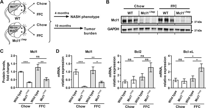
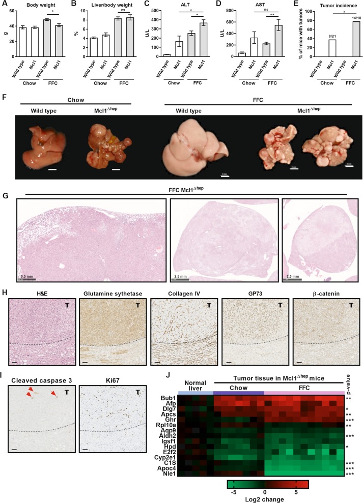
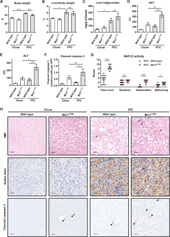
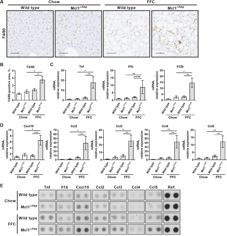
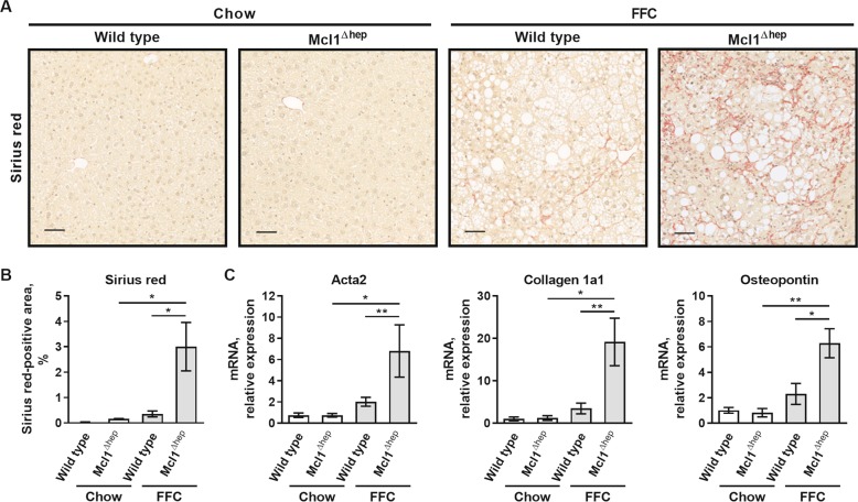
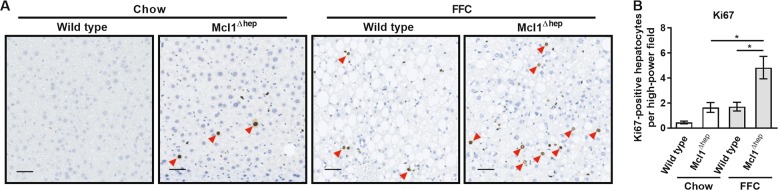
Nonalcoholic fatty liver disease is the most common chronic liver disease and may progress to nonalcoholic steatohepatitis (NASH) and hepatocellular carcinoma (HCC). The molecular determinants of this pathogenic progression, however, remain largely undefined. Since liver tumorigenesis is driven by apoptosis, we examined the effect of overt hepatocyte apoptosis in a mouse model of NASH using mice lacking myeloid cell leukemia 1 (Mcl1), a pro-survival member of the BCL-2 protein family. Hepatocyte-specific Mcl1 knockout (Mcl1) mice and control littermates were fed chow or FFC (high saturated fat, fructose, and cholesterol) diet, which induces NASH, for 4 and 10 months. Thereafter, liver injury, inflammation, fibrosis, and tumor development were evaluated biochemically and histologically. Mcl1 mice fed with the FFC diet for 4 months displayed a marked increase in liver injury, hepatocyte apoptosis, hepatocyte proliferation, macrophage-associated liver inflammation, and pericellular fibrosis in contrast to chow-fed Mcl1 and FFC diet-fed Mcl1-expressing littermates. After 10 months of feeding, 78% of FFC diet-fed Mcl1 mice developed liver tumors compared to 38% of chow-fed mice of the same genotype. Tumors in FFC diet-fed Mcl1 mice were characterized by cytologic atypia, altered liver architecture, immunopositivity for glutamine synthetase, and histologically qualified as HCC. In conclusion, this study provides evidence that excessive hepatocyte apoptosis exacerbates the NASH phenotype with enhancement of tumorigenesis in mice.
非酒精性脂肪性肝病是最常见的慢性肝病,可能进展为非酒精性脂肪性肝炎(NASH)和肝细胞癌(HCC)。然而,这种致病进展的分子决定因素在很大程度上仍未得到明确。由于肝肿瘤发生是由细胞凋亡驱动的,我们使用缺乏髓样细胞白血病 1(Mcl1)的小鼠模型(BCL-2 蛋白家族的一种生存促进成员)检查了 NASH 小鼠中明显的肝细胞凋亡的影响。肝细胞特异性 Mcl1 敲除(Mcl1)小鼠和对照同窝仔鼠分别用普通饲料或 FFC(高饱和脂肪、果糖和胆固醇)饮食喂养 4 个月和 10 个月,后者可诱导 NASH。此后,通过生化和组织学方法评估肝损伤、炎症、纤维化和肿瘤发展。与普通饲料喂养的 Mcl1 和 FFC 饮食喂养的 Mcl1 表达同窝仔鼠相比,用 FFC 饮食喂养 4 个月的 Mcl1 小鼠显示出明显的肝损伤、肝细胞凋亡、肝细胞增殖、巨噬细胞相关的肝炎症和细胞周纤维化增加。喂养 10 个月后,78%的 FFC 饮食喂养的 Mcl1 小鼠与相同基因型的普通饲料喂养的小鼠相比,发展为肝肿瘤。FFC 饮食喂养的 Mcl1 小鼠的肿瘤具有细胞学异型性、改变的肝结构、谷氨酰胺合成酶免疫阳性和组织学上符合 HCC 的特点。总之,这项研究提供了证据表明,过度的肝细胞凋亡加剧了 NASH 表型,并增强了小鼠的肿瘤发生。