Kim Bo-Guen, Song Ju Yeun, Zo Sungmin, Im Yunjoo, Choi Sangjoon, Han Joungho, Jeong Byeong-Ho, Kim Hojoong
Division of Pulmonary and Critical Care Medicine, Samsung Medical Center, Sungkyunkwan University School of Medicine, Seoul, South Korea.
Department of Pathology and Translational Genomics, Samsung Medical Center, Sungkyunkwan University School of Medicine, Seoul, Republic of Korea.
Respir Med Case Rep. 2020 Jan 22;29:101002. doi: 10.1016/j.rmcr.2020.101002. eCollection 2020.
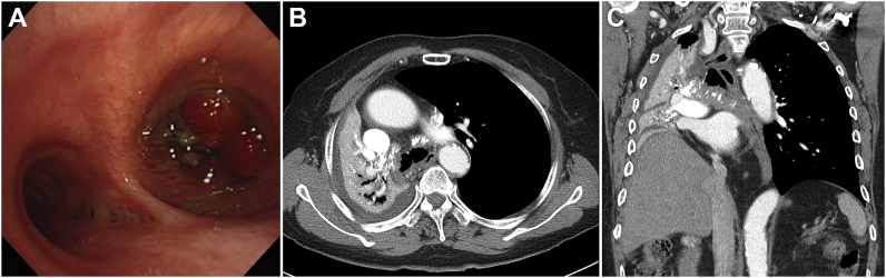
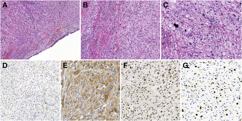
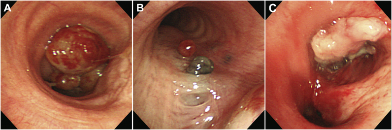
Malignant pulmonary granular cell tumor (GCT) is extremely rare and difficult to distinguish from benign GCT. Most GCTs are neural-type and express S-100. However, a small subset of tumors sub-classified as the non-neural type do not express S-100. We report a case of malignant non-neural-type GCT in the lungs. A 77-year-old woman felt chest discomfort and dyspnea in July 2019. She had never smoked and had no medical history other than hypertension and diabetes mellitus. She was initially evaluated at a local hospital. Flexible bronchoscopy showed total occlusion of the right main bronchus by a mass-like lesion. Biopsy of the mass lesion revealed chronic inflammation. The patient visited for re-evaluation in September 2019. Rigid bronchoscopy showed worsening of the total obstruction of the right main bronchus by a tumor mass, such that the carina was not visible. Additionally, endobronchial nodules were observed on the medial side of left main bronchus. The tumor masses of both main bronchi were removed by bronchoscopic intervention, but the right main bronchus was not opened. Biopsy revealed malignant GCT, favoring the non-neuronal type (S-100-negative). We report an extremely rare case of malignant pulmonary GCT negative for S-100 in immunohistochemistry. In this case, surgical resection was not possible because the tumor was diagnosed at a fairly advanced stage and had spread to involve the contralateral main bronchus. The patient chose to be treated at another hospital and was thereafter lost to follow-up.
恶性肺颗粒细胞瘤(GCT)极为罕见,且难以与良性GCT区分。大多数GCT为神经型,表达S-100。然而,一小部分被归类为非神经型的肿瘤不表达S-100。我们报告一例肺部恶性非神经型GCT病例。一名77岁女性在2019年7月感到胸部不适和呼吸困难。她从不吸烟,除高血压和糖尿病外无其他病史。她最初在当地医院接受评估。柔性支气管镜检查显示右主支气管被一肿块样病变完全阻塞。肿块病变活检显示为慢性炎症。该患者于2019年9月前来复诊。硬质支气管镜检查显示肿瘤肿块使右主支气管完全阻塞情况恶化,以至于隆突不可见。此外,在左主支气管内侧观察到支气管内结节。通过支气管镜介入切除了两个主支气管的肿瘤肿块,但右主支气管未打开。活检显示为恶性GCT,倾向于非神经元型(S-100阴性)。我们报告一例免疫组化S-100阴性的极为罕见的恶性肺GCT病例。在该病例中,由于肿瘤在相当晚期被诊断出且已扩散至对侧主支气管,无法进行手术切除。患者选择到另一家医院治疗,此后失访。