• 文献检索
  • 文档翻译
  • 深度研究
  • 学术资讯
  • Suppr Zotero 插件Zotero 插件
  • 邀请有礼
  • 套餐&价格
  • 历史记录
应用&插件
Suppr Zotero 插件Zotero 插件浏览器插件Mac 客户端Windows 客户端微信小程序
定价
高级版会员购买积分包购买API积分包
服务
文献检索文档翻译深度研究API 文档MCP 服务
关于我们
关于 Suppr公司介绍联系我们用户协议隐私条款
关注我们

Suppr 超能文献

核心技术专利:CN118964589B侵权必究
粤ICP备2023148730 号-1Suppr @ 2026

文献检索

告别复杂PubMed语法,用中文像聊天一样搜索,搜遍4000万医学文献。AI智能推荐,让科研检索更轻松。

立即免费搜索

文件翻译

保留排版,准确专业,支持PDF/Word/PPT等文件格式,支持 12+语言互译。

免费翻译文档

深度研究

AI帮你快速写综述,25分钟生成高质量综述,智能提取关键信息,辅助科研写作。

立即免费体验

鉴定头颈部转移性恶性黑色素瘤中的癌症干细胞亚群。

Identification of Cancer Stem Cell Subpopulations in Head and Neck Metastatic Malignant Melanoma.

机构信息

Gillies McIndoe Research Institute, Newtown, Wellington 6021, New Zealand.

Biostatistical Group/Dean's Department, University of Otago, Wellington 6242, New Zealand.

出版信息

Cells. 2020 Jan 30;9(2):324. doi: 10.3390/cells9020324.

DOI:10.3390/cells9020324
PMID:32019273
原文链接:https://pmc.ncbi.nlm.nih.gov/articles/PMC7072148/
Abstract

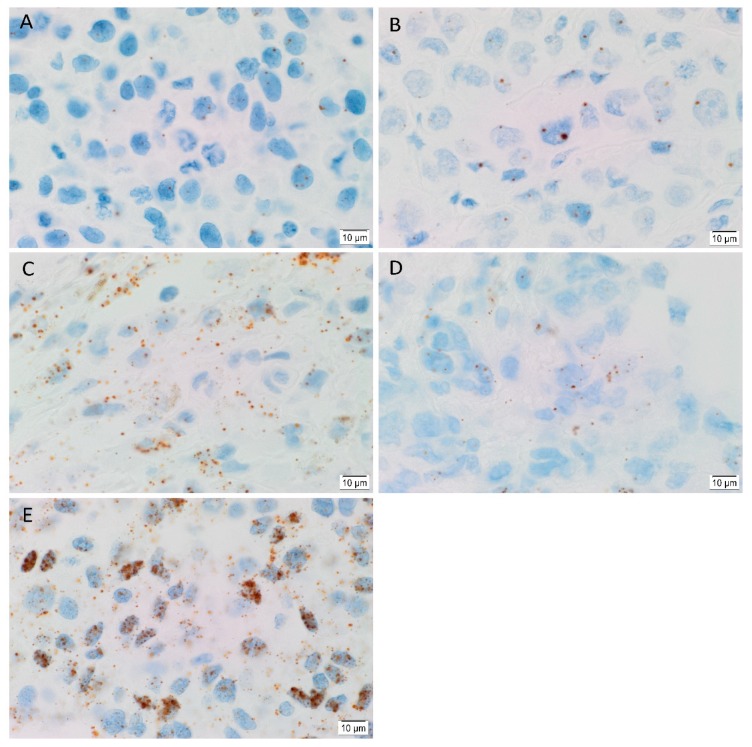
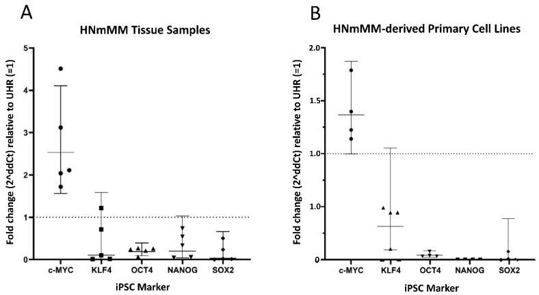
Cancer stem cells (CSCs) have been identified in many cancer types. This study identified and characterized CSCs in head and neck metastatic malignant melanoma (HNmMM) to regional lymph nodes using induced pluripotent stem cell (iPSC) markers. Immunohistochemical (IHC) staining performed on 20 HNmMM tissue samples demonstrated expression of iPSC markers OCT4, SOX2, KLF4, and c-MYC in all samples, while NANOG was expressed at low levels in two samples. Immunofluorescence (IF) staining demonstrated an OCT4+/SOX2+/KLF4+/c-MYC+ CSC subpopulation within the tumor nests (TNs) and another within the peritumoral stroma (PTS) of HNmMM tissues. IF also showed expression of NANOG by some OCT4+/SOX2+/KLF4+/c-MYC+ cells within the TNs in an HNmMM tissue sample that expressed NANOG on IHC staining. In situ hybridization ( = 6) and reverse-transcription quantitative polymerase chain reaction ( = 5) on the HNmMM samples confirmed expression of all five iPSC markers. Western blotting of primary cell lines derived from four of the 20 HNmMM tissue samples showed expression of SOX2, KLF4, and c-MYC but not OCT4 and NANOG, and three of these cell lines formed tumorspheres in vitro. We demonstrate the presence of two putative CSC subpopulations within HNmMM, which may be a novel therapeutic target in the treatment of this aggressive cancer.

摘要

癌症干细胞 (CSCs) 已在许多癌症类型中被鉴定出来。本研究使用诱导多能干细胞 (iPSC) 标志物鉴定并表征了头颈部转移性恶性黑色素瘤 (HNmMM) 至区域淋巴结中的 CSCs。对 20 个 HNmMM 组织样本进行免疫组织化学 (IHC) 染色,结果显示所有样本均表达 iPSC 标志物 OCT4、SOX2、KLF4 和 c-MYC,而 NANOG 在两个样本中低表达。免疫荧光 (IF) 染色显示 HNmMM 组织中肿瘤巢 (TN) 内存在 OCT4+/SOX2+/KLF4+/c-MYC+ 的 CSC 亚群,另一个存在于肿瘤周围基质 (PTS) 内。IF 还显示了在一个表达 NANOG 的 HNmMM 组织样本中,TN 内的一些 OCT4+/SOX2+/KLF4+/c-MYC+细胞表达 NANOG。对 HNmMM 样本进行的原位杂交 (= 6) 和逆转录定量聚合酶链反应 (= 5) 证实了所有五个 iPSC 标志物的表达。对 20 个 HNmMM 组织样本中的 4 个衍生的原代细胞系进行 Western blot 分析显示 SOX2、KLF4 和 c-MYC 的表达,但 OCT4 和 NANOG 的表达则不然,其中 3 个细胞系在体外形成肿瘤球体。我们证明了 HNmMM 中存在两个潜在的 CSC 亚群,这可能是治疗这种侵袭性癌症的新的治疗靶点。

https://cdn.ncbi.nlm.nih.gov/pmc/blobs/c613/7072148/ddaaa97ea270/cells-09-00324-g008.jpg
https://cdn.ncbi.nlm.nih.gov/pmc/blobs/c613/7072148/cb1b4d3d86a9/cells-09-00324-g001.jpg
https://cdn.ncbi.nlm.nih.gov/pmc/blobs/c613/7072148/6190e115498a/cells-09-00324-g002.jpg
https://cdn.ncbi.nlm.nih.gov/pmc/blobs/c613/7072148/c9e30f46c1d3/cells-09-00324-g003.jpg
https://cdn.ncbi.nlm.nih.gov/pmc/blobs/c613/7072148/7f6f0a3b672d/cells-09-00324-g004.jpg
https://cdn.ncbi.nlm.nih.gov/pmc/blobs/c613/7072148/ce9abd149cf9/cells-09-00324-g005.jpg
https://cdn.ncbi.nlm.nih.gov/pmc/blobs/c613/7072148/77445f472a3b/cells-09-00324-g006.jpg
https://cdn.ncbi.nlm.nih.gov/pmc/blobs/c613/7072148/c2c6ce86e733/cells-09-00324-g007.jpg
https://cdn.ncbi.nlm.nih.gov/pmc/blobs/c613/7072148/ddaaa97ea270/cells-09-00324-g008.jpg

相似文献

1
Identification of Cancer Stem Cell Subpopulations in Head and Neck Metastatic Malignant Melanoma.鉴定头颈部转移性恶性黑色素瘤中的癌症干细胞亚群。
Cells. 2020 Jan 30;9(2):324. doi: 10.3390/cells9020324.
2
Cancer Stem Cell Subpopulations Are Present Within Metastatic Head and Neck Cutaneous Squamous Cell Carcinoma.癌症干细胞亚群存在于转移性头颈部皮肤鳞状细胞癌中。
Front Oncol. 2020 Jul 30;10:1091. doi: 10.3389/fonc.2020.01091. eCollection 2020.
3
Cancer stem cell subpopulations in moderately differentiated head and neck cutaneous squamous cell carcinoma.中度分化的头颈部皮肤鳞状细胞癌中的癌症干细胞亚群
Heliyon. 2019 Aug 16;5(8):e02257. doi: 10.1016/j.heliyon.2019.e02257. eCollection 2019 Aug.
4
Cancer Stem Cells in Head and Neck Metastatic Malignant Melanoma Express Components of the Renin-Angiotensin System.头颈部转移性恶性黑色素瘤中的癌症干细胞表达肾素-血管紧张素系统的成分。
Life (Basel). 2020 Nov 2;10(11):268. doi: 10.3390/life10110268.
5
Expression of cathepsins B and D by cancer stem cells in head and neck metastatic malignant melanoma.头颈部转移性恶性黑色素瘤中肿瘤干细胞表达组织蛋白酶 B 和 D。
Melanoma Res. 2021 Oct 1;31(5):426-438. doi: 10.1097/CMR.0000000000000752.
6
Cancer stem cells within moderately differentiated head and neck cutaneous squamous cell carcinoma express components of the renin-angiotensin system.中等分化的头颈部皮肤鳞状细胞癌中的癌症干细胞表达肾素-血管紧张素系统的成分。
J Plast Reconstr Aesthet Surg. 2019 Sep;72(9):1484-1493. doi: 10.1016/j.bjps.2018.11.013. Epub 2018 Nov 22.
7
Colon adenocarcinoma-derived cells that express induced-pluripotent stem cell markers possess stem cell function.表达诱导多能干细胞标志物的结肠腺癌衍生细胞具有干细胞功能。
PLoS One. 2020 May 19;15(5):e0232934. doi: 10.1371/journal.pone.0232934. eCollection 2020.
8
Expression of cancer stem cell markers in metastatic melanoma to the brain.癌症干细胞标志物在脑转移性黑色素瘤中的表达。
J Clin Neurosci. 2019 Feb;60:112-116. doi: 10.1016/j.jocn.2018.10.068. Epub 2018 Oct 24.
9
Cell Populations Expressing Stemness-Associated Markers in Lung Adenocarcinoma.肺腺癌中表达干性相关标志物的细胞群体。
Life (Basel). 2021 Oct 18;11(10):1106. doi: 10.3390/life11101106.
10
Cancer Stem Cells in Metastatic Head and Neck Cutaneous Squamous Cell Carcinoma Express Components of the Renin-Angiotensin System.转移性头颈部皮肤鳞状细胞癌中的癌症干细胞表达肾素-血管紧张素系统的成分。
Cells. 2021 Jan 27;10(2):243. doi: 10.3390/cells10020243.

引用本文的文献

1
Cancer Stem Cells in Melanoma: Drivers of Tumor Plasticity and Emerging Therapeutic Strategies.黑色素瘤中的癌症干细胞:肿瘤可塑性的驱动因素及新兴治疗策略
Int J Mol Sci. 2025 Aug 1;26(15):7419. doi: 10.3390/ijms26157419.
2
Cancer Stem Cells and the Renin-Angiotensin System in the Tumor Microenvironment of Melanoma: Implications on Current Therapies.黑色素瘤肿瘤微环境中的癌症干细胞与肾素-血管紧张素系统:对当前治疗的启示
Int J Mol Sci. 2025 Feb 6;26(3):1389. doi: 10.3390/ijms26031389.
3
EHMT2 Suppresses ARRB1 Transcription and Activates the Hedgehog Signaling to Promote Malignant Phenotype and Stem Cell Property in Oral Squamous Cell Carcinoma.

本文引用的文献

1
Cancer stem cell subpopulations in primary colon adenocarcinoma.原发性结肠腺癌中的癌症干细胞亚群。
PLoS One. 2019 Sep 6;14(9):e0221963. doi: 10.1371/journal.pone.0221963. eCollection 2019.
2
Cancer stem cell subpopulations in moderately differentiated head and neck cutaneous squamous cell carcinoma.中度分化的头颈部皮肤鳞状细胞癌中的癌症干细胞亚群
Heliyon. 2019 Aug 16;5(8):e02257. doi: 10.1016/j.heliyon.2019.e02257. eCollection 2019 Aug.
3
Expression of cancer stem cell markers in metastatic melanoma to the brain.癌症干细胞标志物在脑转移性黑色素瘤中的表达。
EHMT2抑制ARR B1转录并激活刺猬信号通路,以促进口腔鳞状细胞癌的恶性表型和干细胞特性。
Mol Biotechnol. 2025 Apr;67(4):1446-1462. doi: 10.1007/s12033-024-01130-9. Epub 2024 Apr 4.
4
Regulation and signaling pathways in cancer stem cells: implications for targeted therapy for cancer.癌症干细胞中的调控和信号通路:对癌症靶向治疗的启示。
Mol Cancer. 2023 Oct 18;22(1):172. doi: 10.1186/s12943-023-01877-w.
5
CXCR2 expression during melanoma tumorigenesis controls transcriptional programs that facilitate tumor growth.CXCR2 在黑色素瘤发生过程中的表达控制着促进肿瘤生长的转录程序。
Mol Cancer. 2023 Jun 3;22(1):92. doi: 10.1186/s12943-023-01789-9.
6
Cancer Metastasis and Treatment Resistance: Mechanistic Insights and Therapeutic Targeting of Cancer Stem Cells and the Tumor Microenvironment.癌症转移与治疗抗性:癌症干细胞及肿瘤微环境的机制洞察与治疗靶点
Biomedicines. 2022 Nov 21;10(11):2988. doi: 10.3390/biomedicines10112988.
7
Insights Into Vascular Anomalies, Cancer, and Fibroproliferative Conditions: The Role of Stem Cells and the Renin-Angiotensin System.血管异常、癌症和纤维增生性疾病的见解:干细胞和肾素-血管紧张素系统的作用
Front Surg. 2022 Apr 27;9:868187. doi: 10.3389/fsurg.2022.868187. eCollection 2022.
8
Oral Cancer Stem Cells: Therapeutic Implications and Challenges.口腔癌干细胞:治疗意义与挑战
Front Oral Health. 2021 Jul 21;2:685236. doi: 10.3389/froh.2021.685236. eCollection 2021.
9
The Role of Genetic Pathways in the Development of Chemoradiation Resistance in Nasopharyngeal Carcinoma (NPC) Patients.遗传途径在鼻咽癌(NPC)患者放化疗抵抗中的作用。
Genes (Basel). 2021 Nov 21;12(11):1835. doi: 10.3390/genes12111835.
10
Cathepsins B, D, and G Are Expressed in Metastatic Head and Neck Cutaneous Squamous Cell Carcinoma.组织蛋白酶B、D和G在转移性头颈部皮肤鳞状细胞癌中表达。
Front Oncol. 2021 Sep 21;11:690460. doi: 10.3389/fonc.2021.690460. eCollection 2021.
J Clin Neurosci. 2019 Feb;60:112-116. doi: 10.1016/j.jocn.2018.10.068. Epub 2018 Oct 24.
4
OCT4, SOX2, and NANOG positive expression correlates with poor differentiation, advanced disease stages, and worse overall survival in HER2 breast cancer patients.OCT4、SOX2和NANOG的阳性表达与HER2阳性乳腺癌患者的低分化、疾病晚期以及较差的总生存期相关。
Onco Targets Ther. 2018 Nov 6;11:7873-7881. doi: 10.2147/OTT.S173522. eCollection 2018.
5
Characterization of Cancer Stem Cells in Colon Adenocarcinoma Metastasis to the Liver.结肠癌肝转移中癌症干细胞的特征分析
Front Surg. 2018 Jan 22;4:76. doi: 10.3389/fsurg.2017.00076. eCollection 2017.
6
Correlation of Cancer Stem-Cell Markers OCT4, SOX2, and NANOG with Clinicopathological Features and Prognosis in Operative Patients with Rectal Cancer.癌症干细胞标志物OCT4、SOX2和NANOG与直肠癌手术患者临床病理特征及预后的相关性
Yonsei Med J. 2018 Jan;59(1):35-42. doi: 10.3349/ymj.2018.59.1.35.
7
Subcellular localisation of the stem cell markers OCT4, SOX2, NANOG, KLF4 and c-MYC in cancer: a review.癌症中干细胞标志物OCT4、SOX2、NANOG、KLF4和c-MYC的亚细胞定位:综述
J Clin Pathol. 2018 Jan;71(1):88-91. doi: 10.1136/jclinpath-2017-204815. Epub 2017 Nov 27.
8
Intratumor and Intertumor Heterogeneity in Melanoma.黑色素瘤的肿瘤内和肿瘤间异质性
Transl Oncol. 2017 Dec;10(6):956-975. doi: 10.1016/j.tranon.2017.09.007. Epub 2017 Oct 24.
9
Expression of embryonic stem cell markers in acute myeloid leukemia.急性髓系白血病中胚胎干细胞标志物的表达
Tumour Biol. 2017 Jul;39(7):1010428317716629. doi: 10.1177/1010428317716629.
10
Cancer Stem Cells in Oral Cavity Squamous Cell Carcinoma: A Review.口腔鳞状细胞癌中的癌症干细胞:综述
Front Oncol. 2017 Jun 2;7:112. doi: 10.3389/fonc.2017.00112. eCollection 2017.