Yang Fan, Zhang Jiaming, Yang Hua
Department of Thyroid and Breast Surgery, The Central Hospital of Wuhan, Tongji Medical College, Huazhong University of Science and Technology, Wuhan, China,
Onco Targets Ther. 2018 Nov 6;11:7873-7881. doi: 10.2147/OTT.S173522. eCollection 2018.
This study aimed to evaluate the correlations of expression of OCT4, SOX2, and NANOG with clinicopathological features and overall survival (OS) in human epidermal growth factor receptor 2-positive (HER2) breast cancer (BC) patients.
One hundred and thirty-four surgical HER2 BC patients who received doxorubicin and cyclophosphamide followed by paclitaxel and trastuzumab adjuvant therapy were enrolled in this study. Immunofluorescence assay was used to detect OCT4, SOX2, and NANOG expressions. The median follow-up duration was 104 months, and the last follow-up date was December 31, 2017.
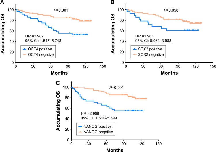
The expressions of OCT4 (=0.001), SOX2 (=0.003), and NANOG (=0.005) were higher in tumor tissues compared with paired adjacent tissues. OCT4 positive expression was associated with poor pathological differentiation (=0.028), larger tumor size (=0.022), advanced N stage (<0.001), and higher TNM stage (<0.001). SOX2 positive expression was correlated with poor pathological differentiation (=0.005), larger tumor size (=0.013), and increased T stage (=0.024). NANOG positive expression was associated with poor pathological differentiation (=0.028), higher N stage (=0.001), and elevated TNM stage (=0.001). Kaplan-Meier curves disclosed that OCT4 (=0.001) and NANOG (=0.001) positive expressions were associated with worse OS, while SOX2 (=0.058) positive expression was only numerically correlated with poor OS, but without statistical significance. Further analyses revealed that co-expression of these three biomarkers disclosed even better predictive value for shorter OS.
OCT4, SOX2, and NANOG positive expressions correlate with poor differentiation and advanced disease stage, and OCT4 and NANOG present with predictive values for poor OS in HER2 BC patients.
本研究旨在评估OCT4、SOX2和NANOG的表达与人类表皮生长因子受体2阳性(HER2)乳腺癌(BC)患者临床病理特征及总生存期(OS)的相关性。
本研究纳入了134例接受多柔比星和环磷酰胺治疗后序贯紫杉醇和曲妥珠单抗辅助治疗的HER2 BC手术患者。采用免疫荧光分析法检测OCT4、SOX2和NANOG的表达。中位随访时间为104个月,最后随访日期为2017年12月31日。
与配对的癌旁组织相比,肿瘤组织中OCT4(P = 0.001)、SOX2(P = 0.003)和NANOG(P = 0.005)的表达更高。OCT4阳性表达与病理分化差(P = 0.028)、肿瘤体积大(P = 0.022)、N分期高(P < 0.001)及TNM分期高(P < 0.001)相关。SOX2阳性表达与病理分化差(P = 0.005)、肿瘤体积大(P = 0.013)及T分期增加(P = 0.024)相关。NANOG阳性表达与病理分化差(P = 0.028)、N分期高(P = 0.001)及TNM分期高(P = 0.001)相关。Kaplan-Meier曲线显示,OCT4(P = 0.001)和NANOG(P = 0.001)阳性表达与较差的OS相关,而SOX2(P = 0.058)阳性表达仅在数值上与较差的OS相关,但无统计学意义。进一步分析显示,这三种生物标志物的共表达对较短的OS具有更好的预测价值。
OCT4、SOX2和NANOG阳性表达与分化差及疾病晚期相关,且OCT4和NANOG对HER2 BC患者的不良OS具有预测价值。