Zhou Lingqi, Tang Hai, Wang Fang, Ou Shanshan, Wu Tong, Fang Yinchao, Xu Jie, Guo Kaihua
Department of Anatomy and Neurobiology, Guangdong Province Key Laboratory of Brain Function and Disease, Zhongshan School of Medicine, Sun Yat‑sen University, Guangzhou, Guangdong 510080, P.R. China.
Guangdong Jiangmen Chinese Medical College, Jiangmen, Guangdong 529000, P.R. China.
Oncol Rep. 2020 Mar;43(3):807-816. doi: 10.3892/or.2020.7459. Epub 2020 Jan 13.
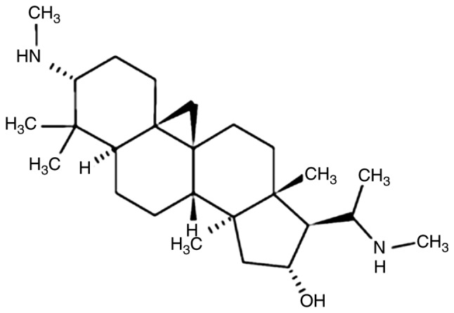
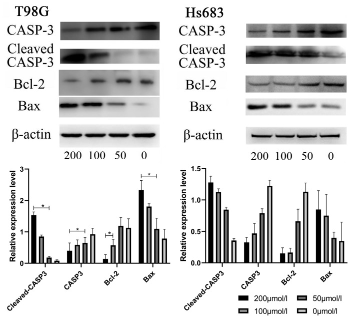
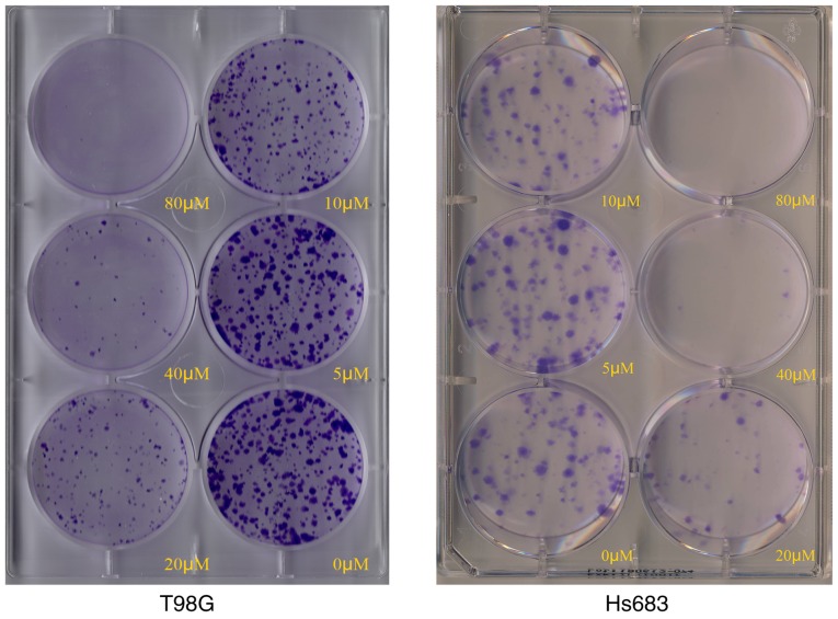
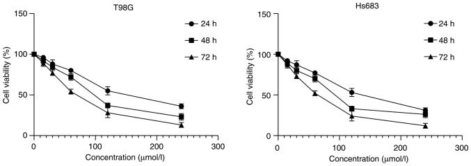
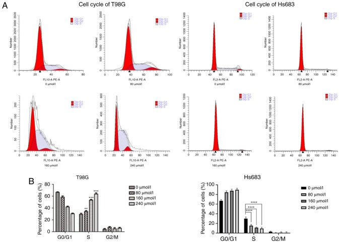
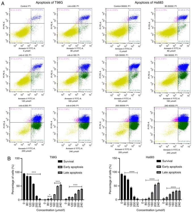
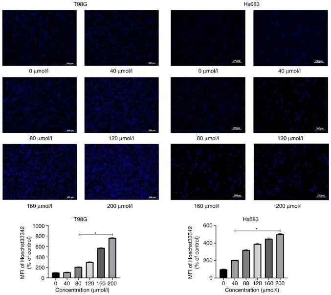
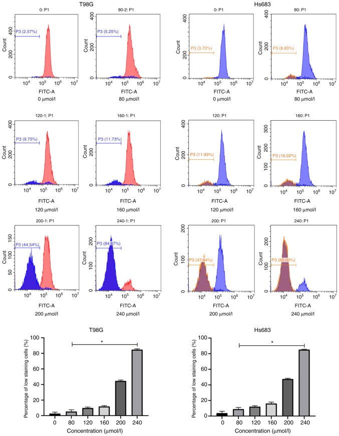
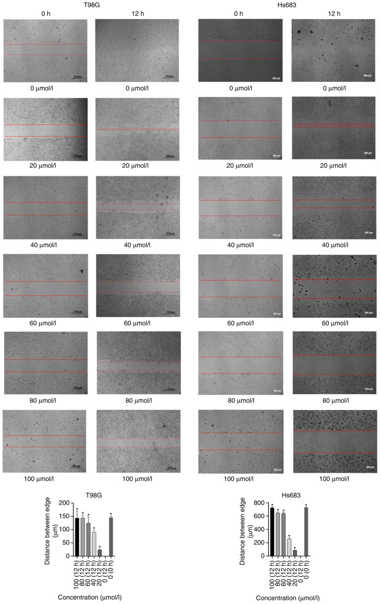
Gliomas are the most common neoplasm of the human central nervous system. Glioblastoma multiforme (GBM) is one of the most serious types of gliomas. Although considerable progress has been made in the development of cancer therapeutic agents, several antineoplastic drugs fail to penetrate the blood‑brain barrier (BBB), resulting in a low survival rate of glioma patients. Recent studies have revealed that the traditional Chinese medicine Buxus microphylla contains the main active component Cyclovirobuxine D (CVB‑D), which can cross the BBB with a novel delivery system. However, it remains unclear whether CVB‑D exerts anticancer effects against GBM and low‑grade glioma (LGG). The aim of the present study was to explore the feasibility of CVB‑D as a new effective agent in the treatment of GBM and LGG. The ability of CVB‑D to inhibit GBM and LGG cell proliferation was detected by CCK8 assay. Flow cytometry was used to detect cell cycle progression and apoptosis induction by Annexin V‑FITC/PI assay. The expression levels of the apoptosis‑associated proteins, namely cleaved caspase‑3 and Bax/Bcl‑2, were detected by western blot analysis. The mitochondrial membrane potential (ΔΨm) was detected by Rh123 dyed fluorescence micrograph. Hoechst staining was used to observe the morphological characteristics of the apoptotic cells. The scratch test was used to evaluate the migration of GBM and LGG cells. The results indicated that CVB‑D reduced cell viability of T98G and Hs683 cells. Flow cytometry demonstrated that CVB‑D‑treated cells were arrested at the S phase of their cell cycle. The expression levels of the apoptosis‑associated proteins were increased in CVB‑D‑treated cells. Rh123 and Hoechst staining indicated morphological changes and mitochondrial membrane potential changes of the cells undergoing apoptosis. The data confirmed that CVB‑D inhibited cell proliferation by arresting the cell cycle of GBM and LLG cells and that it promoted the induction of cell apoptosis by altering the mitochondrial membrane potential. The findings of the present study indicate the potential value of CVB‑D in the treatment of glioma.
胶质瘤是人类中枢神经系统最常见的肿瘤。多形性胶质母细胞瘤(GBM)是最严重的胶质瘤类型之一。尽管在癌症治疗药物的研发方面取得了相当大的进展,但几种抗肿瘤药物无法穿透血脑屏障(BBB),导致胶质瘤患者的生存率较低。最近的研究表明,传统中药小叶黄杨含有主要活性成分环维黄杨星D(CVB-D),它可以通过一种新型递送系统穿过血脑屏障。然而,CVB-D是否对GBM和低级别胶质瘤(LGG)发挥抗癌作用仍不清楚。本研究的目的是探讨CVB-D作为一种新的有效药物治疗GBM和LGG的可行性。通过CCK8测定法检测CVB-D抑制GBM和LGG细胞增殖的能力。采用流式细胞术通过膜联蛋白V-FITC/PI测定法检测细胞周期进程和凋亡诱导情况。通过蛋白质免疫印迹分析检测凋亡相关蛋白(即裂解的半胱天冬酶-3和Bax/Bcl-2)的表达水平。通过Rh123染色荧光显微镜检测线粒体膜电位(ΔΨm)。采用Hoechst染色观察凋亡细胞的形态特征。划痕试验用于评估GBM和LGG细胞的迁移情况。结果表明,CVB-D降低了T98G和Hs683细胞的活力。流式细胞术表明,经CVB-D处理的细胞停滞在细胞周期的S期。经CVB-D处理的细胞中凋亡相关蛋白的表达水平升高。Rh123和Hoechst染色表明凋亡细胞的形态变化和线粒体膜电位变化。数据证实,CVB-D通过阻滞GBM和LLG细胞的细胞周期来抑制细胞增殖,并通过改变线粒体膜电位促进细胞凋亡的诱导。本研究结果表明CVB-D在胶质瘤治疗中的潜在价值。