Sato Ayako, Kakinuma Sei, Miyoshi Masato, Kamiya Akihide, Tsunoda Tomoyuki, Kaneko Shun, Tsuchiya Jun, Shimizu Taro, Takeichi Eiko, Nitta Sayuri, Kawai-Kitahata Fukiko, Murakawa Miyako, Itsui Yasuhiro, Nakagawa Mina, Azuma Seishin, Koshikawa Naohiko, Seiki Motoharu, Nakauchi Hiromitsu, Asahina Yasuhiro, Watanabe Mamoru
Department of Gastroenterology and Hepatology Tokyo Medical and Dental University Tokyo Japan.
Department of Liver Disease Control Tokyo Medical and Dental University (TMDU) Tokyo Japan.
Hepatol Commun. 2019 Dec 24;4(2):235-254. doi: 10.1002/hep4.1459. eCollection 2020 Feb.
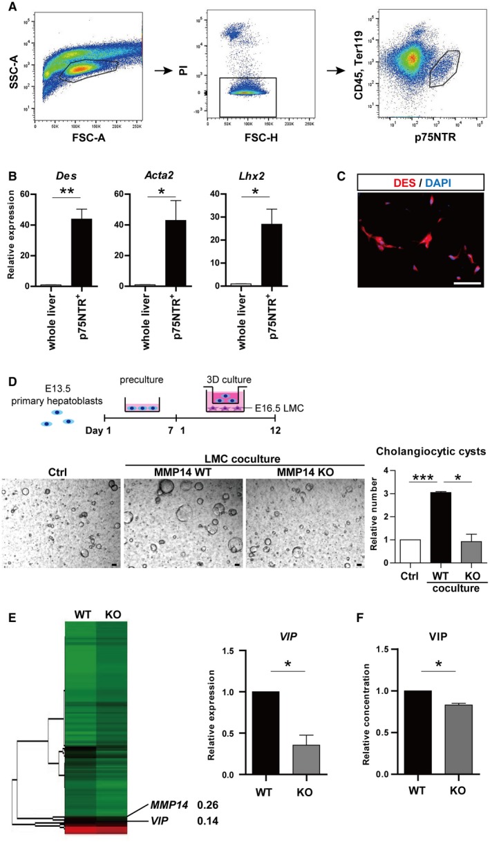
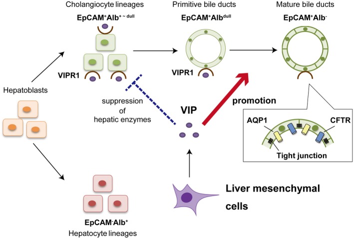
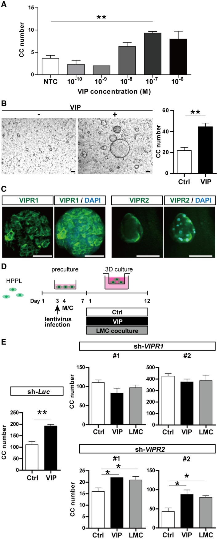
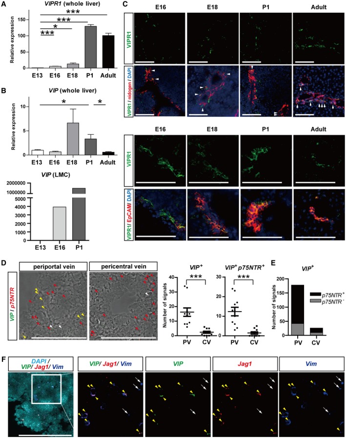
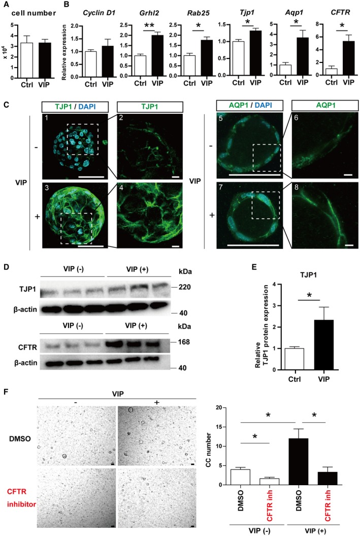
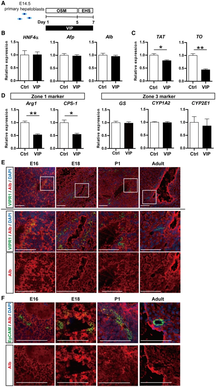
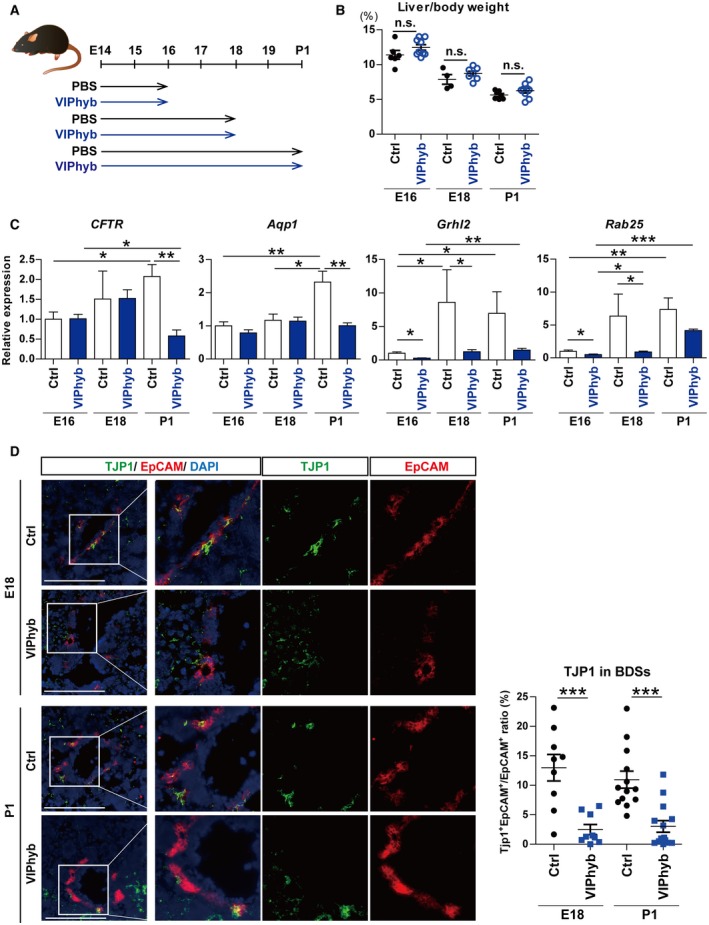
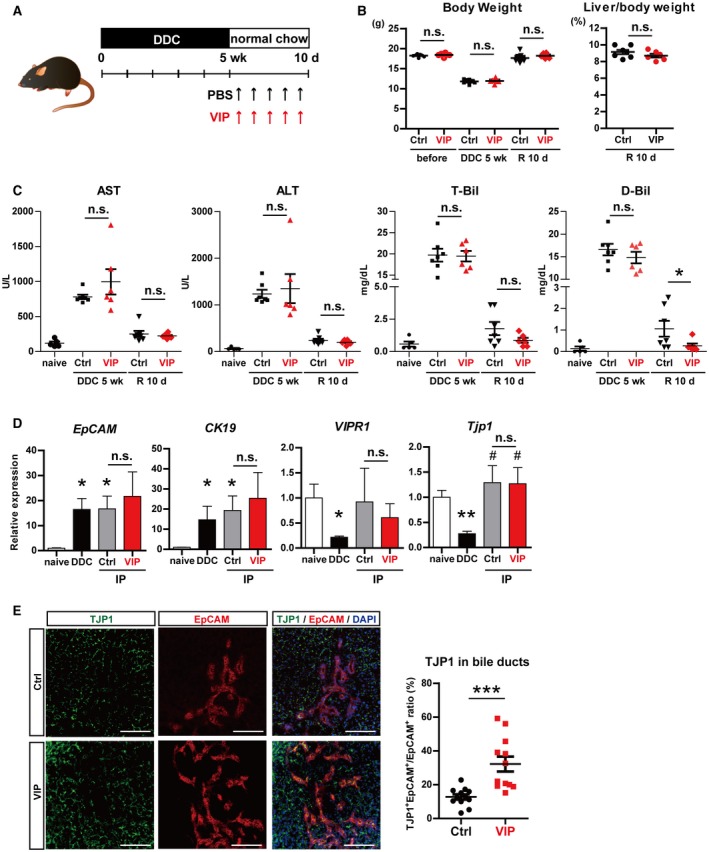
Formation of intrahepatic bile ducts (IHBDs) proceeds in accordance with their microenvironment. Particularly, mesenchymal cells around portal veins regulate the differentiation and ductular morphogenesis of cholangiocytes in the developing liver; however, further studies are needed to fully understand the arrangement of IHBDs into a continuous hierarchical network. This study aims to clarify the interaction between biliary and liver mesenchymal cells during IHBD formation. To identify candidate factors contributing to this cell-cell interaction, mesenchymal cells were isolated from embryonic day 16.5 matrix metalloproteinase 14 (MMP14)-deficient (knockout [KO]) mice livers, in which IHBD formation is retarded, and compared with those of the wild type (WT). WT mesenchymal cells significantly facilitated the formation of luminal structures comprised of hepatoblast-derived cholangiocytes (cholangiocytic cysts), whereas MMP14-KO mesenchymal cells failed to promote cyst formation. Comprehensive analysis revealed that expression of vasoactive intestinal peptide (VIP) was significantly suppressed in MMP14-KO mesenchymal cells. VIP and VIP receptor 1 (VIPR1) were mainly expressed in periportal mesenchymal cells and cholangiocytic progenitors during IHBD development, respectively, . VIP/VIPR1 signaling significantly encouraged cholangiocytic cyst formation and up-regulated tight junction protein 1, cystic fibrosis transmembrane conductance regulator, and aquaporin 1, . VIP antagonist significantly suppressed the tight junction assembly and the up-regulation of ion/water transporters during IHBD development . In a cholestatic injury model of adult mice, exogenous VIP administration promoted the restoration of damaged tight junctions in bile ducts and improved hyperbilirubinemia. VIP is produced by periportal mesenchymal cells during the perinatal stage. It supports bile duct development by establishing tight junctions and up-regulating ion/water transporters in cholangiocytes. VIP contributes to prompt recovery from cholestatic damage through the establishment of tight junctions in the bile ducts.
肝内胆管(IHBDs)的形成与其微环境密切相关。具体而言,门静脉周围的间充质细胞调节发育中肝脏内胆管细胞的分化和胆管形态发生;然而,需要进一步研究以全面了解IHBDs如何排列成连续的分层网络。本研究旨在阐明IHBD形成过程中胆管间充质细胞与肝脏间充质细胞之间的相互作用。为了确定促成这种细胞间相互作用的候选因子,从胚胎第16.5天基质金属蛋白酶14(MMP14)缺陷(敲除[KO])小鼠肝脏中分离间充质细胞,该小鼠肝脏中IHBD形成受阻,并与野生型(WT)小鼠的间充质细胞进行比较。WT间充质细胞显著促进了由肝母细胞衍生的胆管细胞组成的管腔结构(胆管细胞囊肿)的形成,而MMP14-KO间充质细胞未能促进囊肿形成。综合分析显示,MMP14-KO间充质细胞中血管活性肠肽(VIP)的表达显著受到抑制。在IHBD发育过程中,VIP和VIP受体1(VIPR1)分别主要表达于门静脉周围间充质细胞和胆管细胞祖细胞中。VIP/VIPR1信号通路显著促进胆管细胞囊肿形成,并上调紧密连接蛋白1、囊性纤维化跨膜传导调节因子和水通道蛋白1。VIP拮抗剂显著抑制IHBD发育过程中的紧密连接组装以及离子/水转运蛋白的上调。在成年小鼠的胆汁淤积损伤模型中,外源性VIP给药促进了胆管中受损紧密连接的恢复,并改善了高胆红素血症。VIP在围产期由门静脉周围间充质细胞产生。它通过在胆管细胞中建立紧密连接并上调离子/水转运蛋白来支持胆管发育。VIP通过在胆管中建立紧密连接,有助于从胆汁淤积损伤中迅速恢复。