Department of Paediatric Pulmonology and Allergology, Erasmus MC - Sophia Children's Hospital, Rotterdam, The Netherlands.
Department of Radiology and Nuclear Medicine, Erasmus MC, Rotterdam, The Netherlands.
Eur Radiol. 2020 May;30(5):2703-2711. doi: 10.1007/s00330-019-06606-w. Epub 2020 Feb 5.
To estimate airway tapering in control subjects and to assess the usability of tapering as a bronchiectasis biomarker in paediatric populations.
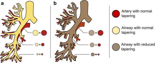
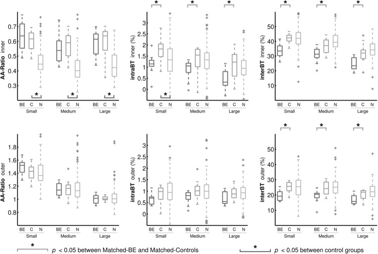
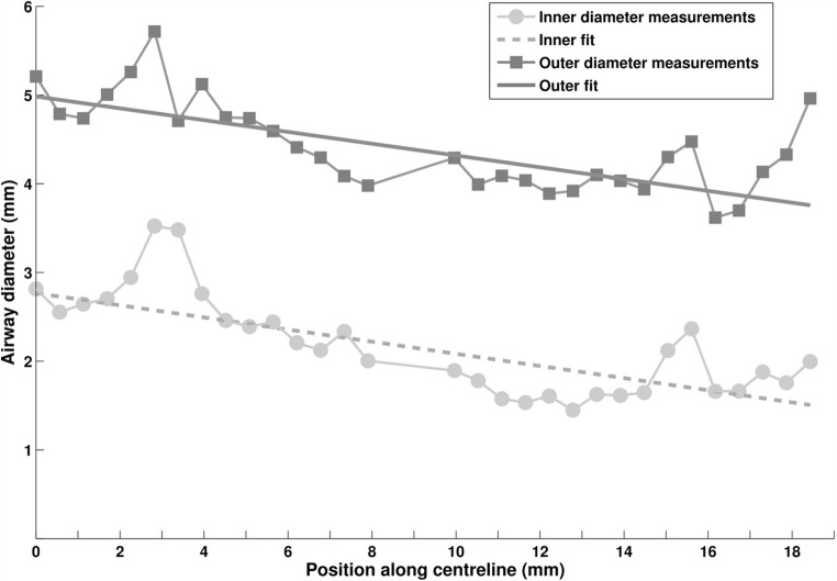
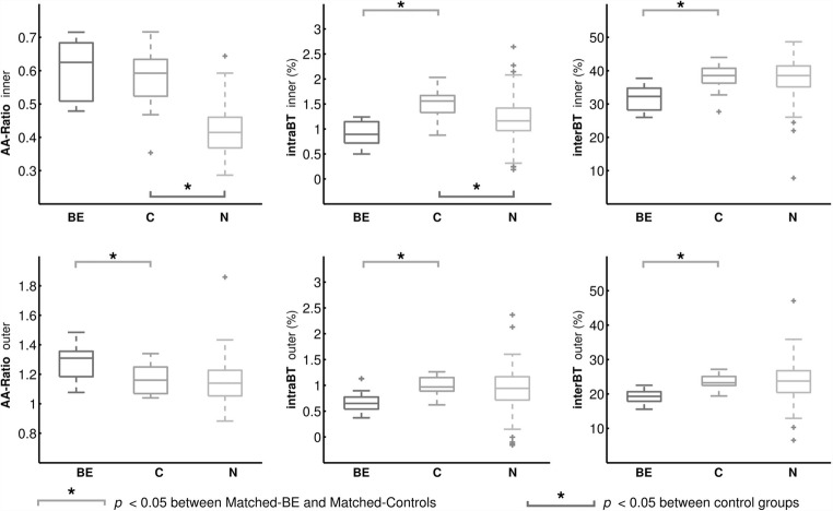
Airway tapering values were semi-automatically quantified in 156 children with control CTs collected in the Normal Chest CT Study Group. Airway tapering as a biomarker for bronchiectasis was assessed on spirometer-guided inspiratory CTs from 12 patients with bronchiectasis and 12 age- and sex-matched controls. Semi-automatic image analysis software was used to quantify intra-branch tapering (reduction in airway diameter along the branch), inter-branch tapering (reduction in airway diameter before and after bifurcation) and airway-artery ratios on chest CTs. Biomarkers were further stratified in small, medium and large airways based on three equal groups of the accompanying vessel size.
Control subjects showed intra-branch tapering of 1% and inter-branch tapering of 24-39%. Subjects with bronchiectasis showed significantly reduced intra-branch of 0.8% and inter-branch tapering of 19-32% and increased airway-artery ratios compared with controls (p < 0.01). Tapering measurements were significantly different between diseased and controls across all airway sizes. Difference in airway-artery ratio was only significant in small airways.
Paediatric normal values for airway tapering were established in control subjects. Tapering showed to be a promising biomarker for bronchiectasis as subjects with bronchiectasis show significantly less airway tapering across all airway sizes compared with controls. Detecting less tapering in larger airways could potentially lead to earlier diagnosis of bronchiectasis. Additionally, compared with the conventional airway-artery ratio, this novel biomarker has the advantage that it does not require pairing with pulmonary arteries.
• Tapering is a promising objective image biomarker for bronchiectasis that can be extracted semi-automatically and has good correlation with validated visual scoring methods. • Less airway tapering was observed in patients with bronchiectasis and can be observed sensitively throughout the bronchial tree, even in the more central airways. • Tapering values seemed to be less influenced by variety in scanning protocols and lung volume making it a more robust biomarker for bronchiectasis detection.
评估对照人群中气道变细的情况,并评估气道变细作为儿童支气管扩张症生物标志物的可用性。
在正常胸部 CT 研究组中,对 156 名患有 CT 对照的儿童进行了气道变细值的半自动定量评估。在 12 名支气管扩张症患者和 12 名年龄和性别匹配的对照患者的肺活量计引导吸气 CT 上评估气道变细作为支气管扩张症的生物标志物。使用半自动图像分析软件定量测量胸部 CT 上的分支内变细(气道直径沿分支减小)、分支间变细(分叉前后气道直径减小)和气道-动脉比。根据伴随血管大小的三个相等组,进一步将生物标志物分为小、中、大气道。
对照受试者表现出 1%的分支内变细和 24-39%的分支间变细。与对照组相比,支气管扩张症患者的分支内变细明显减少(0.8%),分支间变细减少(19-32%),气道-动脉比增加(p<0.01)。在所有气道大小中,疾病患者与对照患者之间的变细测量值均有显著差异。仅在小气道中,气道-动脉比的差异具有统计学意义。
在对照受试者中建立了气道变细的儿科正常值。变细显示出作为支气管扩张症的有前途的生物标志物的潜力,因为与对照组相比,支气管扩张症患者在所有气道大小上的气道变细明显减少。在更大的气道中检测到更少的变细可能会导致支气管扩张症的早期诊断。此外,与传统的气道-动脉比相比,这种新型生物标志物具有不需要与肺动脉配对的优势。
• 变细是一种有前途的支气管扩张症客观图像生物标志物,可以半自动提取,与经过验证的视觉评分方法具有良好的相关性。• 支气管扩张症患者的气道变细减少,并且可以在整个支气管树中敏感地观察到,甚至在更中央的气道中。• 变细值似乎受扫描方案和肺容量变化的影响较小,使其成为检测支气管扩张症的更可靠生物标志物。