Kobe Univ., Japan.
Kyoto Institute of Technology, Japan.
J Biomed Opt. 2020 Feb;25(3):1-15. doi: 10.1117/1.JBO.25.3.032010.
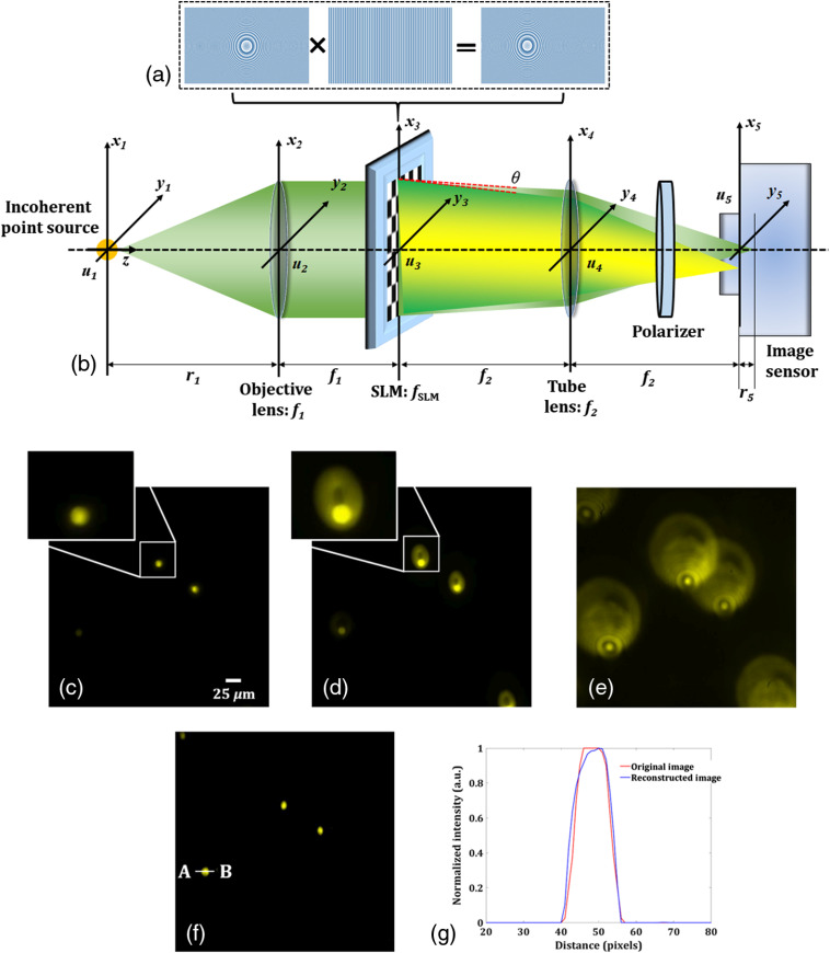
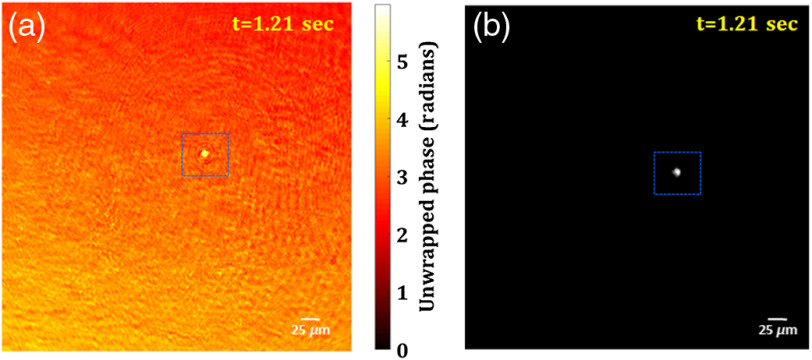
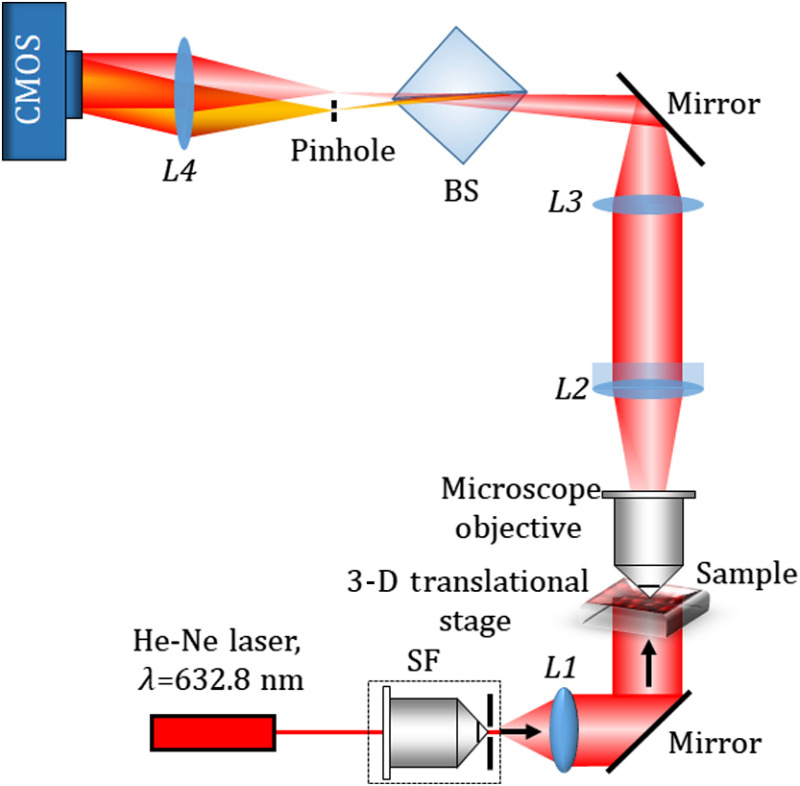
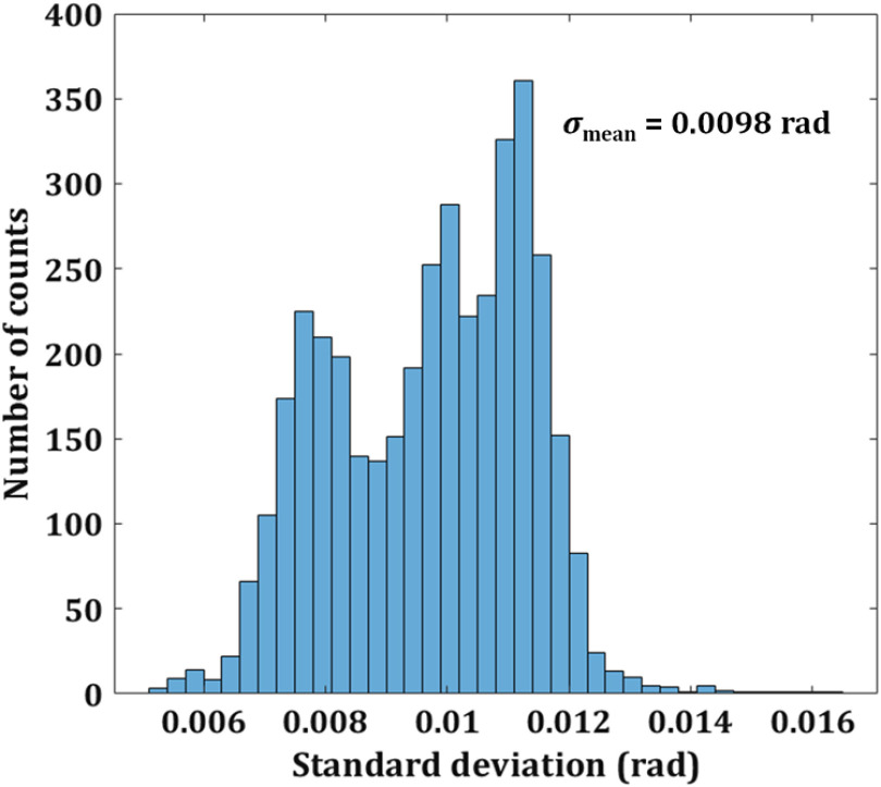
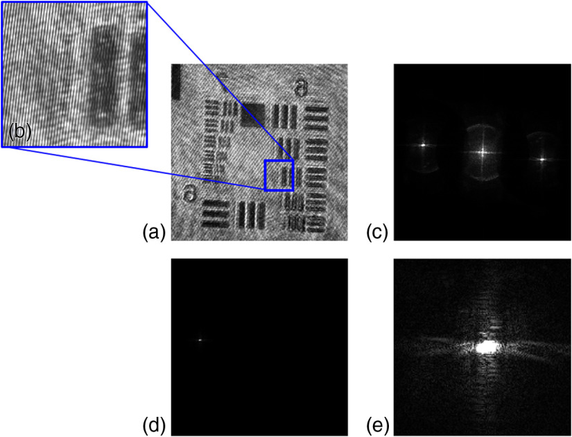
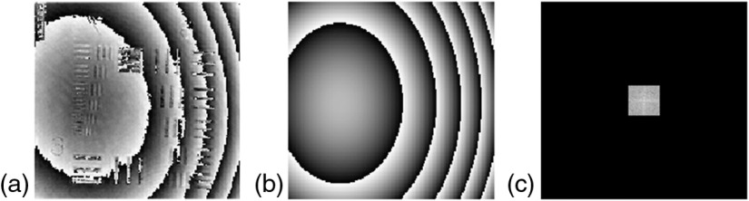
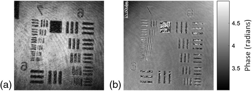
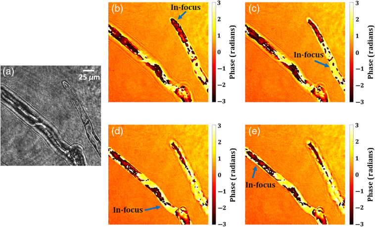
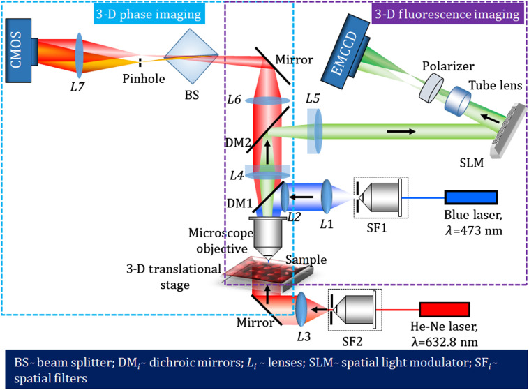
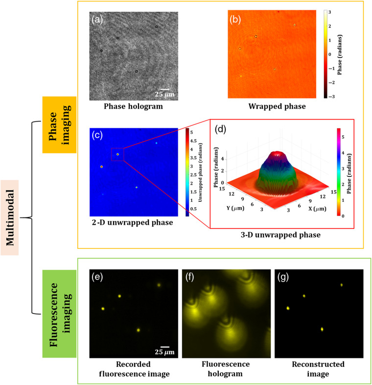
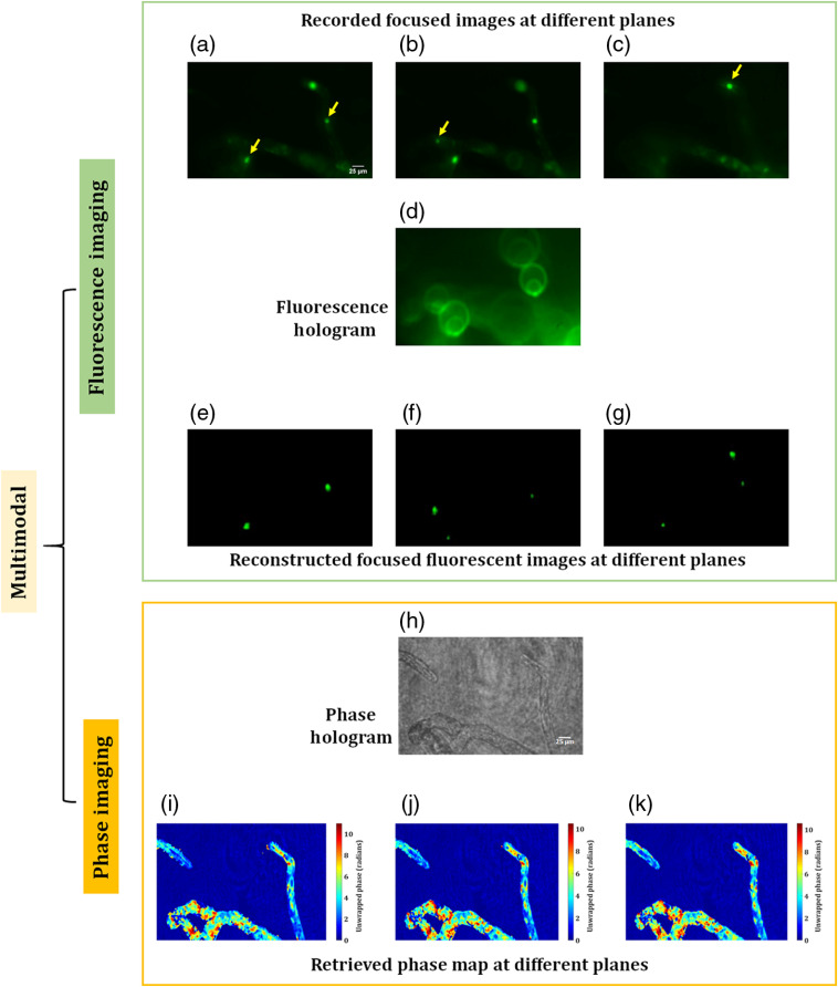
A stable multimodal system is developed by combining two common-path digital holographic microscopes (DHMs): coherent and incoherent, for simultaneous recording and retrieval of three-dimensional (3-D) phase and 3-D fluorescence imaging (FI), respectively, of a biological specimen. The 3-D FI is realized by a single-shot common-path off-axis fluorescent DHM developed recently by our group. In addition, we accomplish, the phase imaging by another single-shot, highly stable common-path off-axis DHM based on a beam splitter. In this DHM configuration, a beam splitter is used to divide the incoming object beam into two beams. One beam serves as the object beam carrying the useful information of the object under study, whereas another beam is spatially filtered at its Fourier plane by using a pinhole and it serves as a reference beam. This DHM setup, owing to a common-path geometry, is less vibration-sensitive and compact, having a similar field of view but with high temporal phase stability in comparison to a two-beam Mach-Zehnder-type DHM. The performance of the proposed common-path DHM and the multimodal system is verified by conducting various experiments on fluorescent microspheres and fluorescent protein-labeled living cells of the moss
通过结合两台常见的共光路数字全息显微镜(DHM):相干和非相干,开发了一个稳定的多模态系统,分别用于同时记录和检索生物样本的三维(3D)相位和 3D 荧光成像(FI)。3D FI 是通过我们小组最近开发的单次共光路离轴荧光 DHM 实现的。此外,我们还通过另一台基于分光镜的单次、高度稳定的共光路离轴 DHM 实现了相位成像。在这种 DHM 配置中,分光镜用于将入射物光束分成两束。一束用作携带研究对象有用信息的物光束,而另一束在其傅里叶平面上通过使用小孔进行空间滤波,用作参考光束。由于共光路几何形状,这种 DHM 装置对振动不敏感,结构紧凑,具有相似的视场,但与双光束马赫-曾德尔型 DHM 相比,具有更高的时间相位稳定性。通过对荧光微球和荧光蛋白标记的苔藓